Learn
Sensitive Teeth: Causes, the Hydrodynamic Mechanism, and Evidence-Based Relief for Dentin Hypersensitivity
Explore why teeth become sensitive: Brännström's hydrodynamic theory, causes of exposed dentin, differential diagnosis, and evidence-based clinical relief.
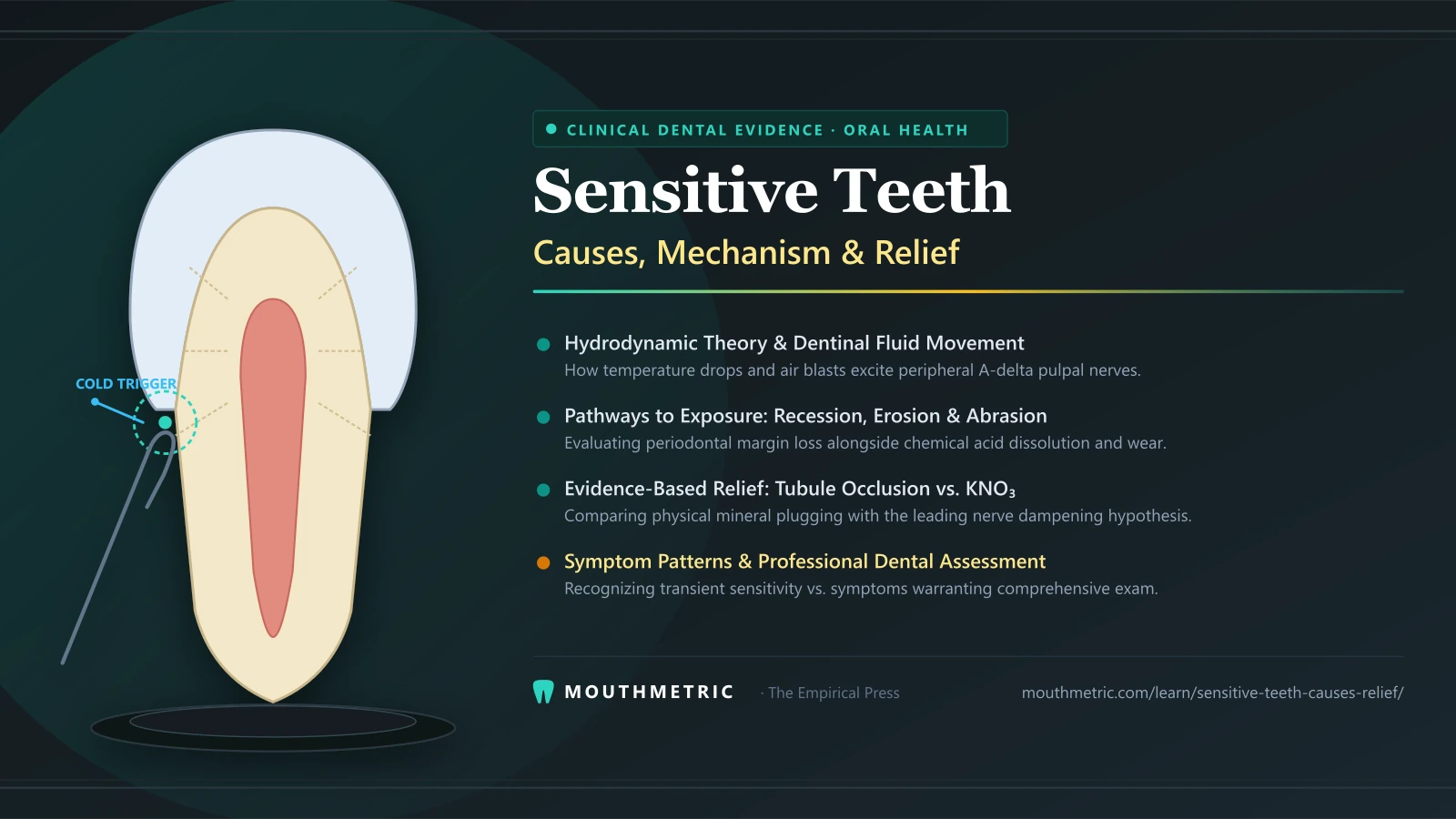
A sudden sip of iced water, a spoonful of hot soup, or even a brisk breath of winter air can trigger a sharp, fleeting jolt through your teeth. For millions of adults, this acute discomfort—commonly referred to as sensitive teeth—is an unsettling daily reality.
When tooth sensitivity strikes, it frequently raises immediate concern. You may wonder whether your enamel is irreversibly damaged, whether a cavity has formed beneath the surface, or whether a root canal procedure is suddenly imminent.
In clinical dentistry, true tooth sensitivity originating from the root or cervical margins is formally classified as dentin hypersensitivity. Unlike the dull, persistent ache of an infected nerve or the structural pain of a cracked tooth, dentin hypersensitivity follows a distinct biophysical mechanism governed by fluid dynamics within microscopic channels inside the tooth.
This guide provides an evidence-based examination of dentin hypersensitivity: what it is according to international consensus, how microscopic fluid movement triggers sharp nerve signals, how receding gums and enamel wear expose sensitive dentin, how to distinguish uncomplicated dentin sensitivity from deeper pulpal or structural conditions, and how active desensitizing ingredients work to provide clinical relief.
Clinical Context & Medical Disclaimer: This article provides independent scientific education and literature review. It does not provide medical or dental advice. Persistent tooth pain, sensitivity lingering after hot stimuli, unprovoked throbbing, or bite tenderness can signal serious endodontic, restorative, or structural conditions that require prompt evaluation by a licensed dental professional.
What Dentin Hypersensitivity Actually Is (and Why It Strikes Suddenly)
To understand tooth sensitivity, it is essential to distinguish between everyday consumer terminology and the clinical condition evaluated in dental practice.
The Consensus Clinical Definition
Dentin hypersensitivity has been standardized by an international consensus of dental researchers and clinical bodies (Canadian Advisory Board on Dentin Hypersensitivity, 2003; Addy, 2002) as:
Short, sharp pain arising from exposed dentin in response to stimuli—typically thermal, evaporative, tactile, osmotic, or chemical—and which cannot be ascribed to any other dental defect or pathology.
This definition establishes three fundamental clinical realities:
- The pain is stimulus-dependent: The sensation arises directly in response to an external trigger (cold air, hot coffee, sweet snacks, or toothbrush bristles).
- The pain is transient: The jolt is sharp and brief, typically dissipating within seconds once the external stimulus is removed.
- It is a diagnosis of exclusion: Dentin hypersensitivity can only be formally diagnosed after a dentist has ruled out other distinct dental pathologies, including dental decay (caries), fractured restorations, cracked teeth, occlusal trauma, and pulpal inflammation (pulpitis).
Epidemiological surveys indicate that dentin hypersensitivity affects between 8% and 57% of the general adult population, with peak prevalence occurring between 20 and 40 years of age (Addy, 2002). Among patients receiving periodontal treatment, prevalence can exceed 60% to 90% due to cervical root exposure.
The Two-Stage Requirement: Dentin Exposure Plus Open Tubules
Dentin is not naturally sensitive when protected by its surrounding biological structures. For dentin hypersensitivity to manifest, dental researchers have established that two sequential conditions must occur:
[Step 1: Lesion Localization]
Gingival recession OR enamel loss exposes the underlying dentin surface.
            │
            ▼
[Step 2: Lesion Initiation]
Acid dissolution OR mechanical wear removes the protective smear layer,
opening (unplugging) the dentinal tubules to the oral environment.
            │
            ▼
[Result: Dentin Hypersensitivity]
External stimuli induce rapid fluid movement, firing pulpal A-delta nerve fibers.- Lesion Localization (Dentin Exposure): The protective covering over dentin must be stripped away. On the tooth crown, this covering is hard dental enamel. Along the cervical neck and root of the tooth, this covering is gingival tissue and a microscopic layer of cementum.
- Lesion Initiation (Tubule Opening): Exposed dentin alone is not necessarily sensitive. Freshly exposed dentin is covered by a natural microscopic film of mineralized cutting debris called the smear layer, and tubule orifices are plugged by mineral deposits. Only when these tubules are opened—rendered patent by dietary acids or abrasive wear—can external forces reach the fluid within.
Inside the Tooth: The Biophysical Mechanism of Dentin Sensitivity
Why does a cold drink cause an instant jolt of pain when touching a tiny patch of exposed root? The answer lies in the microscopic architecture of human dentin and the physics of fluid displacement.
Microscopic Dentin Anatomy: Millions of Fluid-Filled Channels
Underneath the visible enamel crown and outer root cementum sits the bulk of the tooth structure: dentin.
Dentin is a calcified, porous biological tissue traversed by millions of microscopic channels known as dentinal tubules. In a healthy human tooth:
- Dentinal tubules radiate outward from the central dental pulp (where living nerves and blood vessels reside) to the outer enamel-dentin junction or cemento-dentin junction.
- Tubule density ranges from approximately 20,000 tubules per square millimeter near the outer surface to over 45,000 to 50,000 per square millimeter near the dental pulp.
- Each tubule has a microscopic internal diameter of roughly 0.8 to 2.5 micrometers ($0.001\text{–}0.0025\text{ mm}$).
- Every patent tubule is filled with dentinal fluid—an extracellular fluid continuous with the pulpal tissue fluid, containing electrolytes, proteins, and the peripheral cellular extensions (processes) of pulpal cells called odontoblasts.
Brännström’s Hydrodynamic Theory: How Fluid Movement Triggers Pain
For decades, scientists debated how dentin senses pain. Early researchers wondered whether enamel nerves existed (they do not) or whether odontoblast cells acted as direct electrical receptors.
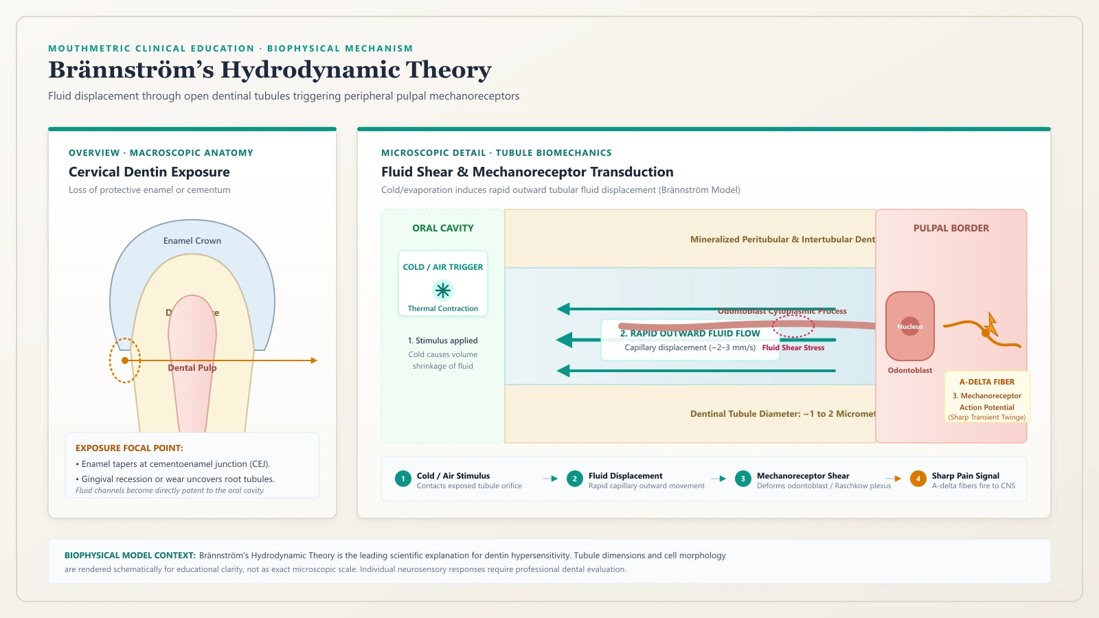
In the 1960s, Swedish dental researcher Mårten Brännström and his colleagues (1964, 1966) introduced and experimentally proved the Hydrodynamic Theory, which remains the universally accepted mechanism for dentin hypersensitivity today.
       [External Stimulus (Cold / Evaporative Air / Osmotic Sugar)]
                                   │
                                   ▼
          [Rapid Outward Fluid Shift in Dentinal Tubules]
                                   │
                                   ▼
        [Mechanical Deformation of Odontoblast Layer & Plexus]
                                   │
                                   ▼
        [Excitation of Mechanosensitive A-Delta Nerve Fibers]
                                   │
                                   ▼
                     [Sharp, Instant Pain Signal]
According to the Hydrodynamic Theory:
- When external stimuli contact open dentinal tubules, they induce a sudden, rapid displacement of the tubular fluid.
- Cold stimuli and evaporative air blasts cause the tubular fluid to contract and pull outward toward the tooth surface at velocities measured in millimeters per second. Because cold causes more rapid fluid movement than warmth, cold is by far the most common and intense trigger of sensitivity.
- Hypertonic osmotic stimuli (such as high-sugar foods or concentrated acidic liquids) draw fluid outward along an osmotic pressure gradient.
- Mechanical tactile stimuli (such as a metal dental explorer or stiff toothbrush filament) physically compress the exposed tubule openings, causing rapid fluid agitation.
- This sudden fluid displacement creates shearing forces and physical pressure changes at the base of the tubule, mechanically deforming the cell membranes of odontoblasts and the unmyelinated nerve endings wrapped around them in the subodontoblastic plexus of Raschkow.
- This mechanical deformation opens stretch-sensitive ion channels, triggering an electrical action potential that travels along sensory nerves to the brain, registered as a sharp, instantaneous flash of pain.
A-Delta vs. C-Fibers: Why Sensitivity Feels Sharp Rather Than Aching
The nature of tooth pain depends entirely on which type of sensory nerve fiber is stimulated within the dental pulp:
| Nerve Fiber Type | Myelination & Conduction Speed | Location in Tooth | Pain Characteristic | Clinical Association |
|---|---|---|---|---|
| A-Delta ($\text{A}\delta$) Fibers | Myelinated; rapid conduction speed ($6\text{–}30\text{ m/s}$) | Concentrated at pulp-dentin border and entering tubule bases | Sharp, bright, lancinating, well-localized | Dentin Hypersensitivity & Reversible Pulpitis |
| C-Fibers | Unmyelinated; slow conduction speed ($0.5\text{–}2\text{ m/s}$) | Distributed deeply throughout the central core of the pulp | Dull, throbbing, aching, diffuse, lingering | Irreversible Pulpitis, Pulpal Necrosis, Abscess |
In dentin hypersensitivity, rapid hydrodynamic fluid displacement selectively excites the mechanosensitive A-delta fibers located at the pulpal periphery. Because A-delta fibers are myelinated and conduct signals rapidly, the pain is perceived immediately as a sharp, brief sting.
In contrast, when bacterial toxins or deep structural inflammation penetrate the central pulp, the slow-conducting C-fibers become activated. C-fiber excitation produces a deep, lingering, throbbing toothache that persists long after any temperature stimulus is gone. Recognizing this biological difference is key to understanding why sensitivity is managed differently from an infected tooth.
How Dentin Becomes Exposed: The Primary Clinical Pathways
Because dentin is sheltered inside the tooth, hypersensitivity cannot occur until the overlying protective biological armor is breached. Dentin exposure occurs through two major clinical pathways.
The Periodontal Pathway: Gingival Recession and Root Exposure
The most common pathway to dentin exposure is the loss of gum tissue height, known as gingival recession.
As detailed in MouthMetric’s clinical guide on Receding Gums: Causes, Measurement, and Treatment, gingival recession occurs when the margin of the gum tissue migrates down the tooth (apically), away from the cementoenamel junction (CEJ) and toward the root apex.
- Along the anatomical root, dentin is not shielded by dense enamel. Instead, it is covered only by a thin, delicate veneer of cementum measuring just $20\text{ to }50,\mu\text{m}$ thick near the gumline.
- Once gingival recession exposes this cervical root surface, the thin cementum layer is easily stripped away within weeks by routine toothbrushing or dissolved by dietary acids.
- Once cementum is lost, the underlying root dentinal tubules are directly exposed to the mouth.
Recession is frequently driven by Periodontal Disease, anatomical tooth position within the dental arch, a thin periodontal tissue phenotype, or prior orthodontic movement.
The Enamel Loss Pathway: Acid Erosion, Tooth Grinding, and Abfraction
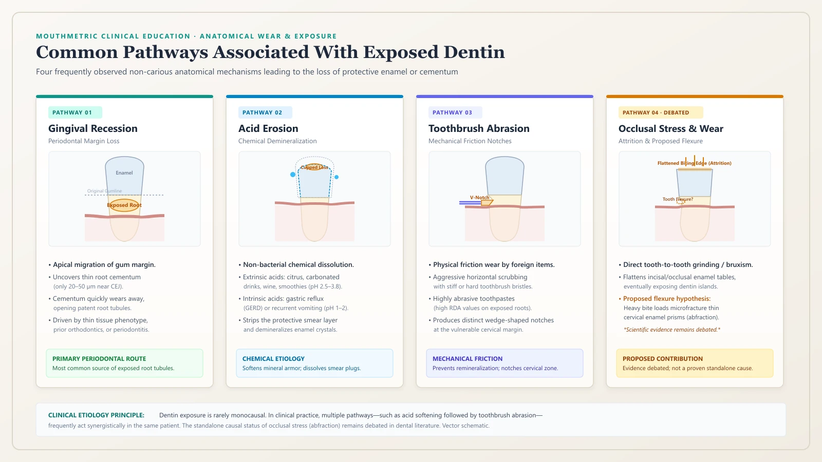
On the anatomical crown of the tooth, dentin is protected by dental enamel—the hardest biological substance in the human body. However, enamel can be gradually worn away through several non-carious wear mechanisms:
- Erosive Tooth Wear (Acid Erosion): Exposure to acids not produced by plaque bacteria.
- Extrinsic sources: Frequent consumption of citrus fruits, carbonated soft drinks, sports beverages, wine, and fruit smoothies (all typically possessing a pH between 2.5 and 3.8).
- Intrinsic sources: Stomach acid (hydrochloric acid, pH 1.0–2.0) contacting the teeth during acid reflux (GERD) or recurrent vomiting. Acid chemically demineralizes and softens the crystalline hydroxyapatite structure of enamel.
- Mechanical Abrasion: The physical wearing away of tooth structure by foreign objects. As evaluated in our guide on Baking Soda and Toothpaste Abrasivity, using toothpastes with high abrasive characteristics on exposed root surfaces or using stiff-bristled toothbrushes with excessive force can accelerate enamel and cementum loss.
- Mechanical Attrition: Wear resulting from direct tooth-to-tooth contact, most commonly caused by nocturnal teeth grinding (bruxism) or clenching. Attrition primarily flattens the biting edges (incisal surfaces) and chewing surfaces (occlusal tables), eventually exposing dentin core islands.
- Abfraction: Wedge-shaped non-carious cervical lesions (NCCLs) occurring near the gumline. Many clinicians consider these lesions to be driven by heavy mechanical bite forces that cause microscopic flexing of the tooth crown, resulting in micro-fractures of the thin cervical enamel prisms near the CEJ.
The Smear Layer: Nature’s Vulnerable Microscopic Seal
When dentin is cut, instrumented, or gently abraded, the mechanical action generates a thin, protective film of crushed mineral crystals and organic matrix fragments known as the smear layer ($1\text{ to }2,\mu\text{m}$ thick).
This smear layer acts as a natural biological sealant: it covers the surface and forms dense microscopic plugs that pack into the mouths of the dentinal tubules. Studies show that an intact smear layer reduces dentin permeability and fluid movement by up to 85% to 90%.
However, the smear layer is acid-labile. Exposure to mild acids—such as salad dressing, fruit juice, or soda—can dissolve these microscopic plugs in less than a minute. Once the smear layer dissolves, the tubular lumens become wide open (patent), and the tooth enters a state of heightened sensitivity.
Sensitivity After Dental Procedures: Bleaching and Deep Cleanings
A frequent point of confusion for patients is experiencing acute sensitivity immediately following professional dental care. Two procedures in particular commonly generate temporary sensitivity: teeth whitening and periodontal deep cleanings.
Teeth Whitening Sensitivity: Peroxide Pulpal Diffusion
As examined in MouthMetric’s comparison of In-Office vs. At-Home Teeth Whitening, post-bleaching sensitivity occurs in approximately 50% to 75% of patients undergoing peroxide-based whitening.
Whitening sensitivity follows a distinct biological pathway that differs fundamentally from classic hydrodynamic tubule flow:
- Hydrogen peroxide ($H_2O_2$) and carbamide peroxide have extremely low molecular weights and easily pass through enamel pores and dentinal tubules.
- Within 15 to 30 minutes of application, peroxide molecules diffuse into the dental pulp chamber.
- In the pulp, free radicals produce transient, mild pulpal irritation (reversible pulpitis) and can directly excite sensory nociceptors—specifically the TRPA1 ion channels on pulpal nerves (Markowitz, 2010).
- This produces sensitivity that can occur spontaneously without any cold or tactile trigger. Bleaching-induced sensitivity is often temporary and self-limiting, commonly subsiding over several days (frequently within 24 to 72 hours in clinical studies) as exposure ends and the transient pulpal irritation resolves.
Post-Scaling and Root Planing Sensitivity: Debridement of the Smear Layer
In periodontal care, patients frequently notice increased root sensitivity following Deep Cleaning Teeth (Scaling and Root Planing).
- During scaling and root planing, ultrasonic and hand instruments thoroughly debride subgingival bacterial biofilm, calculus, and diseased root cementum to arrest periodontitis.
- This mechanical debridement leaves behind freshly exposed root dentin that has had its pre-existing calculus and surface pellicle removed.
- According to European Federation of Periodontology (EFP) S3 clinical practice guidelines (Suvan et al., 2020), transient root sensitivity is a recognized, expected, and self-limiting side effect of subgingival instrumentation.
- In clinical studies, post-scaling sensitivity is commonly reported to peak within the first one to two weeks following treatment and then gradually diminish as salivary minerals form new surface deposits over open tubule orifices, though the timeline varies by individual.
Differential Diagnosis: Dentin Hypersensitivity vs. Other Tooth Pain
Because tooth pain can arise from multiple biological sources, knowing how to interpret symptoms is a vital health literacy skill. Differentiating uncomplicated dentin sensitivity from acute pulpal inflammation or structural tooth damage requires evaluating the character, trigger, and duration of the pain.
                              [Tooth Pain Triggered]
                                        │
        ┌───────────────────────────────┴───────────────────────────────┐
        ▼                                                               ▼
[Stimulus-Dependent Only]                                   [Lingering / Spontaneous]
- Sharp, fleeting jolt                                      - Throbbing or deep ache
- Subsides quickly when trigger removed                     - Lingers or throbs after heat or cold
- Cold, sweet, air, or touch triggers                       - Spontaneous pain at night
        │                                                               │
        ▼                                                               ▼
[Dentin Hypersensitivity                                    [Pulpal / Structural Pathology]
 or Reversible Pulpitis]                                    - Symptomatic Irreversible Pulpitis
                                                            - Pulpal Necrosis / Abscess
                                                            - Cracked Tooth Syndrome
Diagnostic Comparison Matrix
The table below outlines how clinical consensus frameworks (American Association of Endodontists, 2013; Abbott & Yu, 2007) differentiate common dental pain presentations:
| Clinical Feature | Dentin Hypersensitivity | Reversible Pulpitis | Symptomatic Irreversible Pulpitis | Cracked Tooth Syndrome | Dental Caries (Cavity) |
|---|---|---|---|---|---|
| Pain Character | Sharp, brief, lancinating | Sharp, brief, momentary | Dull throbbing, sharp ache, or intense pounding | Sharp, brief twinge on biting or release | Mild twinge to sweet/cold, or silent |
| Primary Triggers | Cold liquids, cold air, sweet/sour, brushing touch | Cold liquids, sweet foods, air blasts | Hot liquids, cold liquids, or entirely spontaneous | Chewing on specific foods (seeds, crusts) | Trapped food, sugar, temperature changes |
| Pain Duration | Fleeting; dissipates shortly after trigger is removed | Brief; dissipates quickly after trigger is removed | Lingers or throbs after trigger is removed | Instant pain upon release of biting pressure | Brief discomfort when food or sugar contacts cavity |
| Spontaneous Pain? | Never | Never | Common; can wake patient from sleep | None unless pulpal involvement has occurred | None until deep dentin or pulp is invaded |
| Heat Response | Rare; occasionally mild brief response | Mild, brief response | Severe, lingering pain; often relieved by cold water | Rare unless crack extends to pulp chamber | Variable |
| Bite Tenderness | None | None | Moderate to severe tenderness on chewing | Severe, sharp pain upon release of bite | None unless structural support fails |
| Primary Origin | Exposed root or worn enamel with open tubules | Mild, reversible inflammation in pulp core | Severe, irreversible inflammation in pulp | Incomplete structural fracture through tooth crown | Bacterial acid demineralization destroying enamel/dentin |
The Duration and Heat Clues: Sharp Transient Twinges vs. Lingering Throbbing
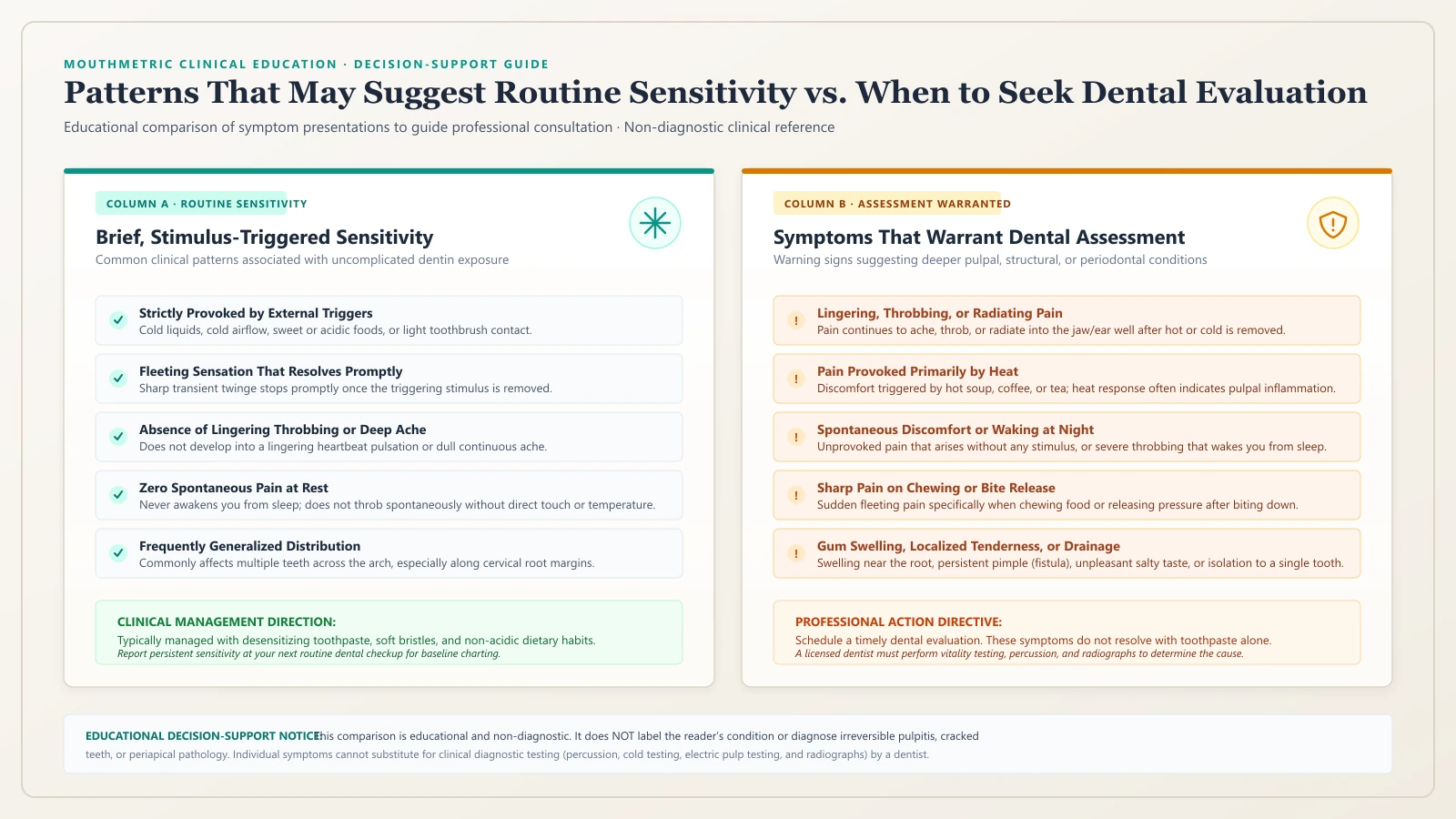
While only a dentist using diagnostic tools (cold testing, electric pulp testing, bite sticks, and periapical radiographs) can definitively diagnose tooth pain, two clinical clues provide critical insight:
- The Duration Clue: In dentin hypersensitivity, pain is tied directly to the physical presence of the stimulus. Once you swallow the cold water or breathe through your nose, hydrodynamic fluid movement stops almost immediately, and the A-delta nerve fibers cease firing. In contrast, if pain lingers, throbs, or radiates well after the cold or hot stimulus is gone, it can be a warning sign that the pulp tissue inside the tooth is actively inflamed and struggling to recover (reversible vs. irreversible pulpitis), requiring professional dental diagnosis.
- The Heat Clue: Dentin hypersensitivity is overwhelmingly triggered by cold, air, or acids. Sensitivity that is triggered primarily by hot drinks (coffee, tea) and results in deep, lingering throbbing is a classic indicator of advanced pulpal inflammation, where elevated temperatures cause expansion of gases and blood vessels in a confined, compromised pulp chamber.
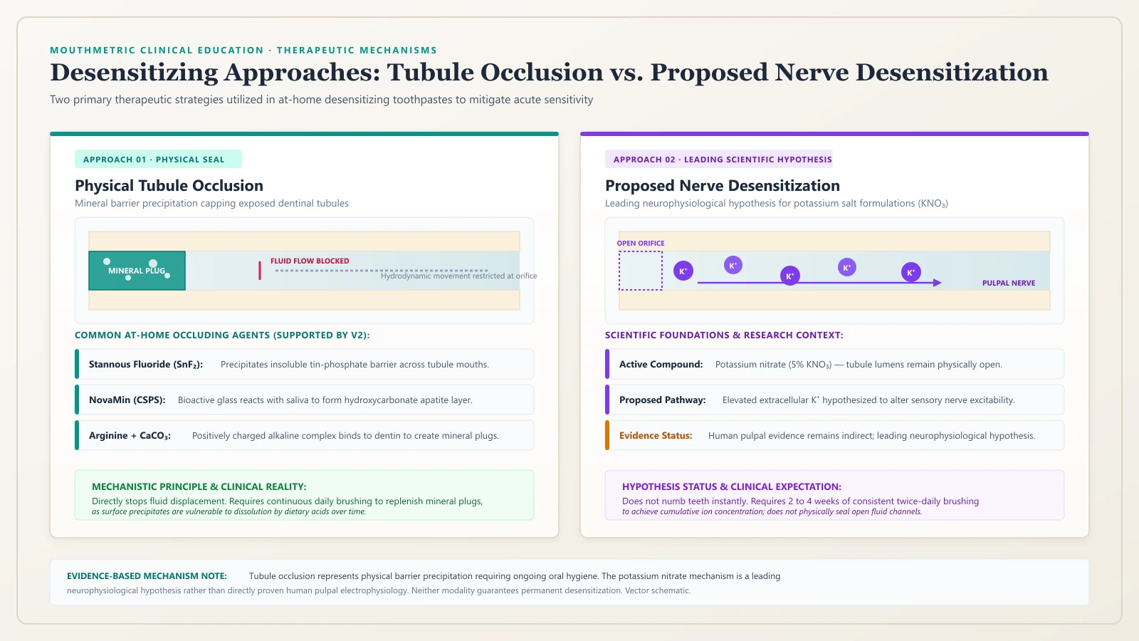
How Desensitizing Treatments Work: The Two Biological Pathways
Managing dentin hypersensitivity relies on two distinct biological strategies: altering the electrical response of the nerve, or physically blocking the microscopic tubules.
                        [Desensitizing Strategies]
                                    │
          ┌─────────────────────────┴─────────────────────────┐
          ▼                                                   ▼
[Pathway 1: Nerve Desensitization (Leading Hypothesis)] [Pathway 2: Tubular Occlusion]
- Potassium Salts (KNO3)                          - Stannous Fluoride (SnF2)
- Leading hypothesis: dampens nerve transmission  - NovaMin / Calcium Sodium Phosphosilicate
- Thought to elevate extracellular K+ ions        - Arginine + Calcium Carbonate
- Aims to reduce action potential firing          - In-Office Varnishes & Resins
- Evaluated over several weeks (commonly 2–8 wks) - Creates physical microcrystalline plugs
Pathway 1: Nerve Depolarization (Potassium Salts)
The first strategy leaves the dentinal tubules completely open but aims to prevent the sensory nerves inside the pulp from sending pain signals to the brain.
- The Active Agent: Potassium Nitrate ($KNO_3$, typically 5%) or potassium chloride.
- The Biological Mechanism: Sensory nerves maintain an electrical resting potential governed by sodium and potassium gradients across their cell membranes. The leading scientific hypothesis—supported by laboratory and electrophysiological research (Markowitz, 2009; Kim, 1986)—proposes that potassium ions ($K^+$) diffuse through open dentinal tubules toward the pulp-dentin junction. In theoretical and animal models, elevating extracellular potassium is thought to depolarize sensory nerve membranes, keeping them in a refractory state that dampens action potential generation. While this neurophysiological mechanism remains the primary proposed explanation, clinical trials focus on its functional outcome: consistent use over several weeks can gradually reduce sensitivity to external triggers.
- Clinical Efficacy & Nuance: A comprehensive Cochrane Systematic Review (Poulsen et al., 2006, PMID: 16855970) evaluated clinical trials on potassium nitrate dentifrices. The meta-analysis confirmed a statistically significant reduction in objective sensitivity scores (tactile sensitivity and cold air blast) after 6 to 8 weeks of use. However, the review also observed that subjective patient-reported relief did not consistently reach statistical significance across all trials, highlighting that individual clinical response varies.
Pathway 2: Physical Tubule Occlusion (Barrier Precipitation)
The second strategy directly targets the physical cause of the problem by creating microscopic mineral plugs or chemical barriers that seal the open tubule mouths, preventing fluid movement entirely.
- The Active Agents: Stannous Fluoride ($SnF_2$), Calcium Sodium Phosphosilicate (NovaMin), Arginine with Calcium Carbonate, and Oxalates.
- The Biological Mechanism: Rather than modifying nerve signaling, these agents deposit insoluble mineral crystals onto the dentin surface and inside the tubular lumens. By physically occluding the tubule openings, they eliminate fluid displacement in response to temperature or touch, stopping hydrodynamic transmission at the source.
Active Ingredient Breakdown: Comparing At-Home Formulations
When reviewing desensitizing toothpastes, consumers encounter several distinct chemical formulations:
| Active Ingredient | Clinical Mechanism | Evidence Strength | Onset Characteristics | Notes & Considerations |
|---|---|---|---|---|
| Potassium Nitrate (5%) | Proposed nerve desensitization; thought to dampen pulpal nerve excitability | Moderate (Cochrane Review Poulsen et al. 2006) | Gradual onset; clinical studies typically evaluate outcomes over several weeks (commonly 2 to 8 weeks) | Does not occlude tubules; symptoms frequently return if discontinued. |
| Stannous Fluoride ($SnF_2$ 0.454%) | Physical occlusion; tin-rich mineral barrier ($Sn_3F_3PO_4$) | Moderate–Strong (Konradsson 2020; Güler 2024) | Moderate onset; builds progressive surface layer | Dual benefit: provides enamel remineralization and anti-gingivitis benefits. |
| Calcium Sodium Phosphosilicate (NovaMin / Bioglass 5%) | Physical occlusion & remineralization; forms hydroxycarbonate apatite | Moderate (Meta-analysis Zhu et al. 2015, PMID: 26544974) | Faster physical occlusion; chemical bonding to dentin | Releases calcium and phosphate in saliva to mimic natural enamel mineral. |
| Arginine (8%) + Calcium Carbonate | Physical occlusion; amino acid binds to dentin, depositing mineral plug | Moderate (Systematic Review Sharif et al. 2013, PMID: 23395975) | Rapid onset demonstrated in multiple clinical trials | Mimics salivary glycoprotein plug formation; resistant to mild acid. |
| Prescription Sodium Fluoride (5,000 ppm NaF) | Enamel remineralization & calcium fluoride globule precipitation | Moderate (ADA Clinical Practice Guidelines) | Progressive over several weeks | High-fluoride prescription; primarily indicated for root caries prevention and adjunct desensitization. |
Clinical Expectations: Relief Timelines and Scientific Evidence
Understanding how desensitizing treatments behave clinically prevents premature frustration and product abandonment.
Why Potassium Nitrate Takes Several Weeks of Daily Use
A common consumer misconception is that brushing once with a sensitive toothpaste will instantly eliminate tooth pain.
Because potassium nitrate relies on the steady diffusion and accumulation of potassium ions through microscopic channels to reach pulpal nerve plexuses, it does not function like an immediate topical numbing gel.
- Clinical trials reviewed in the Cochrane Systematic Review (Poulsen et al., 2006) evaluate outcomes over multi-week intervals (commonly 2, 4, 6, and 8 weeks).
- In practice, relief builds gradually over several weeks of consistent, twice-daily brushing rather than appearing overnight, and individual response varies.
- Furthermore, because potassium salts do not alter tooth structure or seal open tubules, stopping the toothpaste allows salivary washing to gradually deplete extracellular potassium concentrations, frequently causing sensitivity to return within a few weeks.
In-Office Professional Interventions: Varnishes, Primers, and Restorations
When conservative at-home desensitizing toothpastes do not provide sufficient relief over several weeks of regular use—or when symptoms are localized and severe—dental practices provide higher-durability, chairside interventions:
[Level 1: Conservative Chairside Barriers]
Professional 5% Sodium Fluoride Varnish (22,600 ppm F)
Deposits a dense layer of calcium fluoride globules over open tubules.
                           │
                           ▼
[Level 2: Chemical & Biological Sealing]
Glutaraldehyde / HEMA Desensitizing Primers (e.g., Gluma)
Glutaraldehyde cross-links dentinal fluid proteins; HEMA polymerizes within the tubule orifice.
                           │
                           ▼
[Level 3: Adhesive Resin Sealants]
Light-cured dentin bonding resins or glass ionomer coatings
Penetrates the tubule openings to form a polymerized hybrid layer.
                           │
                           ▼
[Level 4: Structural & Surgical Restoration]
Composite restorations (for deep notched NCCL defects)
OR Subepithelial Connective Tissue Grafting (SCTG) for accessible RT1 recession defects.- 5% Sodium Fluoride Varnish (22,600 ppm F): Applied directly to sensitive root surfaces. It forms a dense, immediate precipitate of calcium fluoride globules that mechanically blocks open tubules. While effective immediately, varnishes are gradually worn away over weeks by toothbrushing and require periodic reapplication.
- Glutaraldehyde / HEMA Desensitizers: Solutions containing glutaraldehyde and hydroxyethyl methacrylate (HEMA). Glutaraldehyde causes coagulation and cross-linking of plasma proteins within the dentinal fluid, precipitating biological protein plugs, while HEMA polymerizes inside the tubule collar.
- Resin-Based Dentin Bonding Agents: Light-cured adhesives that infiltrate open tubules and polymerize into microscopic resin tags, forming a long-lasting mechanical seal.
- Cervical Restorations: When dentin loss has formed a deep, notched defect (non-carious cervical lesion) that threatens structural integrity, placing a tooth-colored composite resin or glass ionomer restoration restores anatomical contour and physically seals the exposed dentin surface.
- Periodontal Root Coverage Surgery: As detailed in MouthMetric’s guide to Gum Recession, when sensitivity is driven by visible Cairo RT1 gingival recession, surgical coverage using a subepithelial connective tissue graft (SCTG) can biologically re-cover the exposed root with keratinized gum tissue.
Daily Evidence-Based Habits to Protect Sensitive Teeth
Alongside desensitizing toothpaste, daily hygiene habits play an enormous role in whether dentinal tubules remain sealed or continuously re-opened.
Filament Selection and Brushing Technique
- Use an Ultra-Soft or Soft Toothbrush: Stiff or hard bristles significantly increase mechanical wear on soft root cementum and dentin.
- Adopt a Gentle Modified Bass Technique: Place the brush bristles at a 45-degree angle toward the gumline and use gentle, circular or sweeping motions. Avoid aggressive, horizontal back-and-forth “scrubbing,” which acts like sandpaper against exposed cervical margins.
- The Spit-Don’t-Rinse Rule: After brushing with a desensitizing toothpaste, spit out the excess foam but do not immediately rinse your mouth vigorously with water. Leaving a thin residual film of toothpaste in the mouth allows active potassium ions or occluding minerals to maintain contact with exposed tubules.
Toothpaste Abrasivity: The Role of RDA and Gentle Formulations
Toothpaste abrasivity is measured by the Relative Dentin Abrasivity (RDA) standard. The international safety standard (ISO 11609) and the American Dental Association set the upper ceiling of safety at 250 RDA.
While there is no mandatory regulatory threshold requiring an RDA below 70, clinical in situ research shows that exposed dentin is substantially softer than enamel (Mohs hardness ~3.0 for dentin vs ~5.0 for enamel). For individuals with exposed root surfaces and sensitive teeth:
- Select toothpastes formulated specifically for sensitivity, which generally use low-to-moderate abrasive silica systems.
- Avoid heavily abrasive “whitening” or tartar-control toothpastes that rely on coarse mechanical abrasives to scrub away surface stains, as these can strip the smear layer and exacerbate cervical wear.
Dietary Acid Management: Balancing Remineralization and Hygiene
- Minimize Frequent Acid Contact: Sipping acidic drinks (citrus water, soda, sports drinks, wine) throughout the day causes repeated acid demineralization cycles that continuously dissolve tubule plugs. Consume acidic beverages with meals rather than sipping them over extended hours.
- Rinse with Water After Acidic Food: Immediately after consuming acidic foods or beverages, rinse your mouth thoroughly with plain water to help neutralize acidity and facilitate natural salivary remineralization.
- Avoid Immediate Scrubbing After Severe Acid Exposure: Because strong dietary acids temporarily soften the microscopic outer mineral layer of enamel and exposed dentin, waiting approximately 30 minutes after consuming high-acid foods or drinks before brushing is a sensible precaution to allow saliva to partially buffer acidity, reducing the risk of brushing away acid-softened minerals.
Critical Warning Signs: When Tooth Pain Requires Urgent Care
Uncomplicated dentin hypersensitivity involves exposed dentin without active infection of the dental pulp. However, because its symptoms can overlap with serious dental conditions, you should not rely on sensitive toothpaste if you experience any of the following warning signs:
- Pain That Lingers or Throbs After Temperature Changes: Pain that lingers well after a hot or cold stimulus—especially when severe, throbbing, or provoked by heat—can be concerning for pulpal disease or nerve involvement and should be promptly evaluated by a dentist.
- Pain Triggered by Heat: Pain that is provoked by warm foods or hot coffee and feels relieved by cold water is a classic clinical indicator of late-stage pulpal disease.
- Spontaneous or Nocturnal Pain: Pain that strikes without any temperature trigger or wakes you from sleep indicates active pulpal or periapical infection.
- Pain on Chewing or Releasing a Bite: Sharp, localized pain that occurs when biting down on hard food or when releasing chewing pressure strongly points toward a cracked tooth or broken cusp.
- Localized Gum Swelling or Fistula: A small bump, pimple, or swelling on the gum near a sensitive tooth indicates a periapical or periodontal abscess, representing an infection that requires prompt dental treatment.
Evidence Review & Methodology
At MouthMetric, we evaluate oral health conditions and products through our structured five-label claims framework to separate marketing claims from independent clinical evidence.
The Five-Label Claims Framework
- VERIFIED FACT: Checkable anatomical and physical facts confirmed by reference standards (e.g. human dentinal tubule density ranges between 20,000 and 50,000 tubules/$mm^2$; ISO 11609 establishes the upper safety limit for toothpaste abrasivity at 250 RDA).
- INDEPENDENT EVIDENCE: Findings from peer-reviewed clinical trials, Cochrane systematic reviews, or professional consensus guidelines published by independent research bodies (e.g. Poulsen et al. 2006 Cochrane review on potassium nitrate; Zhu et al. 2015 meta-analysis on calcium sodium phosphosilicate).
- EDITORIAL INTERPRETATION: MouthMetric’s balanced synthesis of the clinical literature (e.g. recognizing that while potassium nitrate reduces objective stimulus sensitivity over 6–8 weeks, subjective patient satisfaction is variable and dependent on continuous daily application).
- USER REPORT: Self-reported patient experiences and consumer survey observations regarding sensitivity triggers or taste preferences.
- VENDOR CLAIM: Statements made by oral care manufacturers regarding “instant relief” or proprietary desensitizing formulations that have not been independently replicated in multi-center clinical trials.
For full details on our editorial standards and conflict-of-interest policies, visit the MouthMetric Methodology.
Frequently Asked Questions About Sensitive Teeth
Can sensitive teeth go away on their own?
Yes, in some cases. If sensitivity is caused by a temporary disruption of the smear layer (such as following a routine cleaning or a mild acid challenge), natural minerals in saliva (calcium and phosphate) can gradually precipitate over several weeks to form new intratubular mineral plugs, naturally dampening sensitivity. However, if root surfaces remain persistently exposed to aggressive brushing or ongoing dietary acids, sensitivity typically persists or recurs without preventive care.
How long does it take for sensitive toothpaste to start working?
It depends on the active ingredient. Toothpastes formulated with potassium nitrate are thought to alter nerve excitability; clinical studies show that relief builds gradually over several weeks of continuous twice-daily use (trials commonly track outcomes between 2 and 8 weeks), though individual response varies. Toothpastes formulated with tubule occluders (such as stannous fluoride, NovaMin, or arginine) can begin forming physical microcrystalline barriers sooner, but maximal relief is generally achieved over several weeks of continuous application.
Why did my teeth become sensitive after a professional cleaning?
During scaling and root planing, dental instruments remove accumulated subgingival calculus and bacterial plaque from root surfaces. While essential for halting periodontal disease, this cleaning process exposes root dentinal tubules that were previously covered by tartar. This post-cleaning sensitivity is common and often temporary—frequently peaking during the first week or two—though some patients may need desensitizing toothpaste or professional varnishes if sensitivity lingers.
Is sensitive teeth a sign of a cavity?
It can be, but they feel different. Dentin hypersensitivity is typically widespread across multiple teeth and triggers a short, sharp jolt that stops immediately when the stimulus is removed. A cavity (dental caries) is usually isolated to a single tooth and may generate pain when sweet food becomes physically trapped inside the defect. If sensitivity is confined to one specific tooth, have it examined by a dentist.
Does teeth whitening permanently ruin tooth enamel or cause permanent sensitivity?
No. High-concentration peroxide whitening gels temporarily increase enamel permeability and diffuse into the pulp, creating mild, transient pulpal excitation. However, clinical studies show that this sensitivity is often temporary and typically resolves over several days (commonly within 24 to 72 hours following the end of bleaching treatments), without causing lasting damage to enamel structure or healthy pulp tissue.
What is the difference between potassium nitrate and stannous fluoride?
They work through completely different biological mechanisms. Potassium nitrate is formulated to act on pulpal nerves: according to the leading neurophysiological hypothesis, potassium ions diffuse along tubules to dampen nerve firing, gradually reducing sensitivity over several weeks of daily use. Stannous fluoride is a tubule occluder: tin and fluoride ions react with dentin minerals to form an insoluble physical plug that caps open tubule mouths, preventing fluid movement from occurring in the first place.
Can brushing too hard cause tooth sensitivity?
Yes. Aggressive brushing with hard bristles or excessive manual pressure—particularly using horizontal sawing motions—can mechanically wear away delicate cervical root cementum and thin enamel prisms. This abrasive wear uncovers dentinal tubules and initiates dentin hypersensitivity.
When should I stop trying sensitive toothpaste and see a dentist?
You should see a dentist if sensitivity does not improve with consistent use of a desensitizing toothpaste, if symptoms worsen or interfere with eating, or immediately if pain lingers after temperature changes, occurs spontaneously, or is felt when chewing.
References
- Canadian Advisory Board on Dentin Hypersensitivity. Consensus-based recommendations for the diagnosis and management of dentin hypersensitivity. J Can Dent Assoc. 2003;69(4):221–226. PMID: 12662460.
- Brännström M, Åström A. A study on the mechanism of pain elicited from the dentin. J Dent Res. 1964;43:619–625. PMID: 14170327. DOI: 10.1177/00220345640430041601.
- Brännström M. Sensitivity of dentine. Oral Surg Oral Med Oral Pathol. 1966;21(4):517–526. PMID: 5218080. DOI: 10.1016/0030-4220(66)90411-7.
- Addy M. Dentine hypersensitivity: New perspectives on an old problem. Int Dent J. 2002;52(S5P2):367–375. DOI: 10.1002/j.1875-595X.2002.tb00936.x.
- Poulsen S, Errboe M, Lescay Mevil Y, Glenny AM. Potassium nitrate toothpaste for dentine hypersensitivity. Cochrane Database Syst Rev. 2006;(3):CD001476. PMID: 16855970. DOI: 10.1002/14651858.CD001476.pub2.
- Markowitz K. The original desensitizers: Strontium and potassium salts. J Clin Dent. 2009;20(5):145–151. PMID: 19902638.
- Zhu M, Li J, Chen B, Mei L, Yao L, Tian J, Li H. The effect of calcium sodium phosphosilicate on dentin hypersensitivity: a systematic review and meta-analysis. PLoS One. 2015;10(11):e0140176. PMID: 26544974. DOI: 10.1371/journal.pone.0140176.
- Sharif MO, Iram S, Brunton PA. Effectiveness of arginine-containing toothpastes in treating dentine hypersensitivity: a systematic review. J Dent. 2013;41(6):483–492. PMID: 23395975. DOI: 10.1016/j.jdent.2013.01.009.
- Konradsson K, Emilson CG, Lingström P, Johannsen G, Ramberg P, Johannsen A. Stabilized stannous fluoride dentifrice in relation to dental caries, dental erosion and dentine hypersensitivity: a systematic review. Am J Dent. 2020;33(2):95–105.
- Markowitz K. Pretty painful: why does tooth bleaching hurt? Med Hypotheses. 2010;74(5):835–840. PMID: 20045265. DOI: 10.1016/j.mehy.2009.11.044.
- American Association of Endodontists (AAE). Endodontic Diagnosis. AAE Colleagues for Excellence, Fall 2013. [Online reference: aae.org].
- Suvan J, Leira Y, Moreno Sancho FM, Graziani F, Derks J, Tomasi C. Subgingival instrumentation for treatment of periodontitis. A systematic review. J Clin Periodontol. 2020;47 Suppl 22:42–75. PMID: 31889320. DOI: 10.1111/jcpe.13245.
- Abbott PV, Yu C. A clinical classification of the status of the pulp and the root canal system. Aust Dent J. 2007;52(1 Suppl):S17–S31. PMID: 17658022. DOI: 10.1111/j.1834-7819.2007.tb00522.x.
- Cobb CM. Clinical significance of subgingival debridement: an evidence-based perspective. J Periodontol. 2002;73(5):560–569. PMID: 12010523. DOI: 10.1902/jop.2002.73.5.560.
- Jaeggi T, Lussi A. Toothbrush abrasion of sound and eroded enamel–an in situ study. Caries Res. 1999;33(6):455–461. PMID: 10529535. DOI: 10.1159/000016551.
- Cummins D. Dentin hypersensitivity: from diagnosis to a breakthrough therapy for everyday sensitivity relief. J Clin Dent. 2009;20(1):1–9. PMID: 19489188.
Sources
- Canadian Advisory Board on Dentin Hypersensitivity (2003) — accessed 2026-10-05
- Brännström M & Åström A (1964) — accessed 2026-10-05
- Brännström M (1966) — accessed 2026-10-05
- Addy M (2002) — accessed 2026-10-05
- Poulsen S et al. (Cochrane Systematic Review, 2006) — accessed 2026-10-05
- Markowitz K (2009) — accessed 2026-10-05
- Zhu M et al. (2015) — accessed 2026-10-05
- Sharif MO et al. (2013) — accessed 2026-10-05
- Markowitz K (2010) — accessed 2026-10-05
- Suvan J et al. (EFP S3 Guideline, 2020) — accessed 2026-10-05
- American Association of Endodontists (2013) — accessed 2026-10-05
- Abbott PV & Yu C (2007) — accessed 2026-10-05