Learn
Gum Disease: Causes, Stages, and Clinical Management
Understand gum disease from gingivitis to periodontitis. Learn what dental pocket numbers mean, causes of bone loss, and evidence-based treatments.
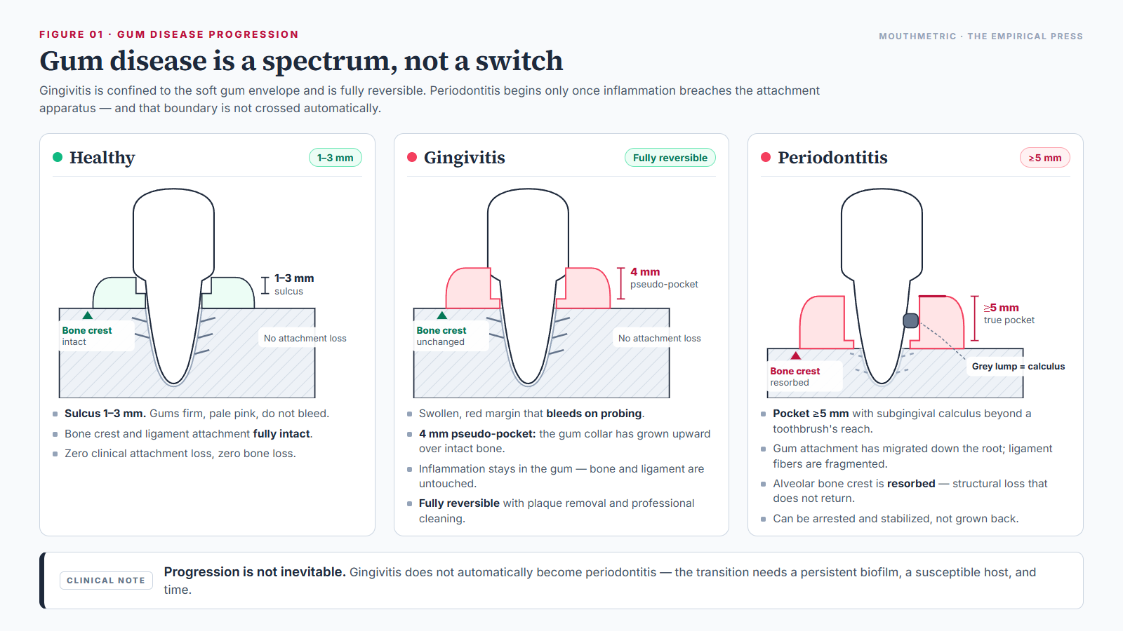
Gum disease — periodontal disease, in clinical language — is a chronic inflammatory condition of the tissues that surround and support the teeth. It is not one condition but a spectrum. At one end sits gingivitis: mild, superficial inflammation of the gum tissue that clears completely once plaque is removed. At the other sits periodontitis: deeper inflammation that has breached the gum seal and damaged the periodontal ligament and the alveolar bone, structure the body does not rebuild.
Gingivitis precedes periodontitis, but the two are not a single track running to one destination. Not all gingivitis progresses. Whether inflammation stays superficial or advances depends on the biofilm, the host response, and the risk factors set out below.
MouthMetric Medical Evidence Review
Clinical, staging, and numeric claims in this article are traced to cited peer-reviewed studies, consensus guidelines, or national health surveys, with full references listed at the end of the page. We do not diagnose, and this page cannot assess your individual situation: pocket depths, bone levels, and tooth stability can only be measured by a licensed dental professional using a calibrated periodontal probe and diagnostic radiographs. Nothing here replaces an examination, a diagnosis, or a treatment plan from your dentist or periodontist, and no passage should be read as a reason to delay care, change a prescription, or treat yourself at home. Periodontal disease is a chronic condition with systemic implications, so decisions about treatment belong in a clinical chair rather than on a web page. For how we select, grade, and update evidence, see the MouthMetric evidence methodology.
What Is Gum Disease?
To understand gum disease, start with the structure it attacks. The periodontium — the biological apparatus anchoring each tooth in the jaw — consists of four specialized tissues:
- Gingiva (the gums): the protective mucosal covering that seals around the neck of the tooth crown and forms the first barrier against oral bacteria.
- Periodontal ligament (PDL): a network of specialized collagen fibers that suspends the tooth root in its socket and absorbs the forces of chewing.
- Cementum: a calcified layer covering the tooth root, giving the ligament fibers an anchorage surface.
- Alveolar bone: the specialized ridge of jawbone forming the tooth sockets.
In health, the gum forms a tight collar around the tooth, and a shallow crevice — the gingival sulcus — sits between the free gum margin and the tooth surface, usually 1 to 3 millimeters deep.
Gum disease begins when bacterial plaque is left undisturbed at and below that gum margin. Clinicians classify the result into two categories:
- Gingival disease (gingivitis): inflammation limited strictly to the gum epithelium and the connective tissue beneath it, with no loss of attachment fibers and no loss of supporting bone.
- Periodontal disease (periodontitis): inflammation that has extended into the deeper structures, degrading the periodontal ligament, root cementum, and alveolar bone.
This is an extremely common chronic condition rather than an unusual infection. Surveys led by Paul Eke and colleagues using the National Health and Nutrition Examination Survey (NHANES) estimate that periodontitis affects roughly 42% to 47% of American adults aged 30 and older — on the order of 64 million people — and an estimated 70% of adults aged 65 and older. Severe periodontitis, the form carrying the greatest risk of tooth loss, affects about 8% to 9% of adults aged 30 and older.
Two things follow from those figures. If you have it, you are in very large company, and there is nothing shameful about it — this is a biofilm-driven inflammatory disease, not a verdict on how well you clean. And severity is a spread: most adults with periodontitis do not have the severe form.
Gingivitis vs. Periodontitis: The Reversibility Boundary
The boundary that matters clinically is the boundary of irreversibility. Gingivitis is confined to the soft gum envelope, so removing the microbial biofilm lets the microvasculature normalize, the cellular infiltrate clear, and the collagen fibers recover. Once the periodontal ligament fibers and the alveolar crest have been degraded, that architectural loss does not return under ordinary conditions.
Harald Löe and colleagues at the Royal Dental College in Aarhus, Denmark, demonstrated the boundary directly in 1965. Systemically healthy volunteers stopped all oral hygiene. Within 10 to 21 days of undisturbed plaque accumulation, every participant developed generalized clinical gingivitis. When normal hygiene was resumed, healthy gums returned in about one week, approximately 7 days. The study established two facts that still govern practice: plaque is the direct cause of gingivitis, and gingivitis resolves completely once plaque is removed.
What makes gingivitis reversible is where it stops. Inflammation confined to the gum epithelium and connective tissue leaves the underlying architecture intact — zero clinical attachment loss and zero radiographic bone loss. Once inflammation breaches that barrier and degrades the ligament fibers and bone crest, the damage is structural, and structural damage is what periodontitis is.
The clinical comparison below synthesizes the diagnostic criteria defined by the 2017 World Workshop classification system.
| Clinical parameter | Gingivitis | Periodontitis |
|---|---|---|
| Primary process | Superficial, plaque-induced inflammation of the gum tissue | Deep, microbially triggered, host-mediated destruction of the attachment apparatus |
| Tissue involved | Gum epithelium and underlying connective tissue only | Gums, periodontal ligament, root cementum, and alveolar bone |
| Clinical attachment loss (CAL) | None attributable to periodontitis | Present — interdental attachment loss of 1 mm or more, depending on stage |
| Alveolar bone loss | None visible on radiographs | Present — measurable crestal resorption on bitewing or periapical radiographs |
| Pocket depth | Normal (3 mm or less), or pseudo-pockets created by swollen tissue | Pathological pockets, typically 4 mm and deeper |
| Bleeding on probing | Bleeding at 10% or more of sites, generalized at 30% or more | Bleeds at active sites, usually alongside deepened pockets |
| Reversibility | Fully reversible with consistent plaque removal and professional cleaning | Structural loss is irreversible; the condition can be arrested and stabilized |
Longitudinal evidence shows that periodontitis is always preceded by gingival inflammation, but gingivitis does not automatically become periodontitis. Gingivitis can persist for years as a stable superficial condition without ever breaching the attachment apparatus. Progression requires a biofilm that has shifted into a destructive dysbiosis, a host susceptible to that shift, and enough uninterrupted time.
How Gum Disease Develops: From Biofilm to Bone Loss
Step 1: Plaque biofilm accumulates and the community shifts
Plaque is a structured biofilm, not a loose layer of germs. Left undisturbed at the gum margin, it matures from a predominantly gram-positive, oxygen-tolerant community of cocci into a gram-negative, anaerobic, rod-dominated community, and it eventually calcifies into dental calculus through mineral precipitation from saliva. Calculus itself is not toxic, but its rough, porous surface is a non-shedding reservoir that keeps sheltering biofilm from the toothbrush.
Step 2: Keystone pathogens push the community into dysbiosis
George Hajishengallis and colleagues proposed in 2012 that a small number of organisms can act as keystone pathogens. Porphyromonas gingivalis is the standard example: present in low abundance, it interferes with host defenses such as complement-mediated bacterial killing, and the whole community reorganizes into a destructive dysbiosis rather than simply overgrowing. The organisms most consistently associated with deep pockets and bleeding are the Red Complex — Porphyromonas gingivalis, Treponema denticola, and Tannerella forsythia — identified by Socransky and colleagues in 1998.
Step 3: The host response causes most of the damage
This is the point most people get wrong. Bacteria trigger the process, but they do not eat bone. The inflammatory response the body mounts to contain them produces the majority of tissue breakdown — the figure usually cited is roughly 80%. Cytokines such as interleukin-1 beta and tumor necrosis factor alpha recruit and activate the cells that dismantle collagen and bone in an attempt to retreat from a bacterial front line the tissue cannot clear.
Step 4: Collagen is cut and bone is resorbed
Infiltrating inflammatory cells release matrix metalloproteinase-8 (MMP-8), a collagen-destroying enzyme that cleaves the collagen fibers of the periodontal ligament. In parallel, inflammatory signaling raises the RANKL to OPG ratio, which switches on osteoclasts and causes them to resorb the alveolar jawbone. Clinically, this is the loss that does not come back.
Step 5: The halitosis link
Deep subgingival pockets are oxygen-poor, which suits anaerobic bacteria that break down proteins in blood and gingival crevicular fluid into volatile sulfur compounds — notably methyl mercaptan (CH₃SH). That is the specific mechanism by which periodontal disease produces a persistent, deep mouth odor that surface freshening does not touch. For the wider range of oral and non-oral causes of malodor, see our guide to the causes of bad breath.
None of these steps is inevitable. Progression from inflammation to destruction needs a persistent biofilm, a susceptible host, and time.
What Causes Gum Disease? Primary Drivers and Risk Modifiers
It is worth separating a primary cause — the agent without which the disease does not start — from a risk modifier, which changes how vulnerable someone is, how fast disease advances, or how severe it becomes.
The primary cause is bacterial plaque biofilm. Plaque that is not disrupted mechanically every day matures, and its calcified form, dental calculus (tartar), creates a rough surface that shelters further subgingival biofilm and cannot be brushed off at home. Plaque alone, however, does not explain why one person with similar hygiene develops only superficial gingivitis while another suffers rapid bone loss. Severity is set by the interaction between the biofilm and the host’s systemic defenses.
Tobacco smoking
In an NHANES III analysis of 12,329 participants, Scott Tomar and Samira Asma (2000) found that current smokers had roughly four times the odds of periodontitis compared with never-smokers, and that more than half — approximately 53% — of adult periodontitis cases in the United States are linked to current or former cigarette smoking. Smoking impairs microvascular flow, creates cellular hypoxia in the gum, suppresses neutrophil function, enriches anaerobic pathogens, and severely blunts healing after treatment. Smoking is the largest modifiable behavioural risk factor for periodontitis, and cessation is the single most valuable change a smoker can make.
Diabetes mellitus
The relationship between diabetes and periodontitis runs in both directions, as documented by Preshaw and colleagues (2012). Hyperglycemia drives the accumulation of advanced glycation end-products (AGEs) in periodontal tissues, which bind their receptors on endothelial cells and macrophages and generate intense local oxidative stress. Poorly controlled diabetes raises the risk of periodontitis roughly threefold. In the other direction, chronic periodontal inflammation contributes to systemic inflammation and worsens glycemic control — and treating periodontitis is associated with an average HbA1c reduction of about 0.3% to 0.4% at three to four months. Gum care and diabetes care are the same conversation.
Genetic susceptibility
Twin studies by Michalowicz and colleagues (2000) indicate that roughly 50% of the variance in periodontitis severity is attributable to genetic factors, while plaque-induced gingivitis shows essentially no heritability. Genetic variation in inflammatory cytokines, pattern-recognition receptors, and antibody subclasses shapes how vigorously a person’s immune system responds to the same biofilm. Genetics creates susceptibility, not destiny: a genetic predisposition changes the effort required, not the possibility, of staying healthy.
Hormonal shifts during pregnancy
A systematic review and meta-analysis by Chen, Hong, and Yu (2022) found that bleeding on probing affects up to 67% of pregnant women, with periodontitis prevalence around 40%. Rising estrogen and progesterone increase capillary permeability and favour organisms such as Prevotella intermedia, producing exaggerated bleeding from tissue that is already inflamed. Pregnancy gingivitis is an amplified inflammatory response to the same plaque; it does not by itself mean accelerated bone loss.
If you are pregnant or planning a pregnancy: periodontal care is not something to postpone. The European Federation of Periodontology review by Bobetsis and colleagues (2020) states that dental practitioners should provide periodontal treatment to pregnant women and that it can be provided safely for both mother and unborn child, with non-surgical treatment generally scheduled during the second trimester. Tell your dental team where you are in the pregnancy and let them plan timing with you; do not delay or skip care on your own advice.
Medication-induced gingival enlargement
Calcium channel blockers such as nifedipine and amlodipine, the anticonvulsant phenytoin, and the immunosuppressant cyclosporine can stimulate gum fibroblast overgrowth, as reviewed by Seymour, Ellis, and Thomason (2000). The resulting overgrowth creates deep pseudo-pockets that trap biofilm and make hygiene difficult even when the patient is careful.
Medication and gums: consult the prescribing physician If your gums are enlarging and you take one of these medicines, raise it with the doctor who prescribes it. Do not stop, skip, or change the dose of a prescribed medicine because of a web page. Separately, medicines that reduce saliva flow — antihistamines, antidepressants, and diuretics among them — remove part of the mouth’s natural cleansing and buffering, which can speed plaque accumulation.
Cardiovascular association, and its limits
Periodontitis is statistically associated with atherosclerotic cardiovascular disease, and the plausibility is not in doubt: the ulcerated pocket wall allows bacteria and bacterial endotoxins into the bloodstream during ordinary chewing, producing low-grade systemic inflammation. What is not established is causation. In a scientific statement for the American Heart Association, Peter Lockhart and a multidisciplinary panel (2012) reviewed the literature and concluded that periodontitis has not been proven to cause atherosclerotic vascular disease, and that periodontal treatment has not been proven to prevent heart attacks, strokes, or other cardiovascular events. Claims that “gum disease causes heart attacks” or that “cleaning your gums protects your heart” go beyond the evidence and should be treated accordingly.
Signs and Symptoms: Why Gum Disease Is Often Silent
The most dangerous property of periodontitis is how quietly it progresses. In the large majority of people, early and moderate disease produces no pain at all, and pain often appears only when destruction is already advanced.
The clinical signs to know about are:
- Bleeding gums. Bleeding during brushing, flossing, or eating is the most common early sign. Inflamed pocket lining becomes micro-ulcerated, and the exposed capillary loops rupture under minimal friction. Healthy tissue does not bleed under ordinary brushing.
- Redness and swelling. Healthy gums are pale coral-pink and firm with a stippled texture. Inflamed margins become smooth, glossy, swollen, and bright red or purplish.
- Recession, or “longer teeth.” As bone and ligament resorb, the gum margin migrates, exposing yellowish root surface and opening triangular dark spaces between teeth.
- Persistent bad breath or a bad taste. Deep pockets harbor anaerobic bacteria that convert proteins into volatile sulfur compounds. If odor persists despite good hygiene, the cause is worth identifying — see our bad breath research hub for the range of recognized causes and what can and cannot be self-assessed.
- Loose teeth or a shifting bite. In advanced disease, reduced support lets teeth move, spread, flare, or develop new gaps.
- Dull ache on chewing. Chronic periodontitis is usually painless, but advanced cases can produce a deep, dull pressure when chewing firm food.
- Pus on gentle pressure. Suppuration from the gum margin indicates an active, deep pocket with a high concentration of inflammatory cells.
Symptoms cannot grade the disease Severity cannot be self-assessed from how things feel. A person with heavily swollen gums may have no structural bone loss at all, while a heavy smoker with pale, non-bleeding gums may already have advanced disease. Only professional probing and radiographic examination can establish where a mouth sits on the spectrum.
Two distinctions keep the picture clear. Superficial bleeding from inflamed gum margins is a sign of gingivitis and is treatable; a tooth that feels loose is a sign of lost support and needs prompt evaluation. And the absence of symptoms means less than it seems: pain is a late and unreliable signal in this disease.
How Dentists Diagnose and Measure Gum Disease
A periodontal diagnosis is a structured, multi-parameter examination performed by a licensed dentist or periodontist, and it requires full-mouth charting together with full-mouth radiographs. The measurements below are clinical procedures; a periodontal probe is not a home instrument, and readers should not attempt to interpret their own readings.
1. Periodontal probing and probing depth
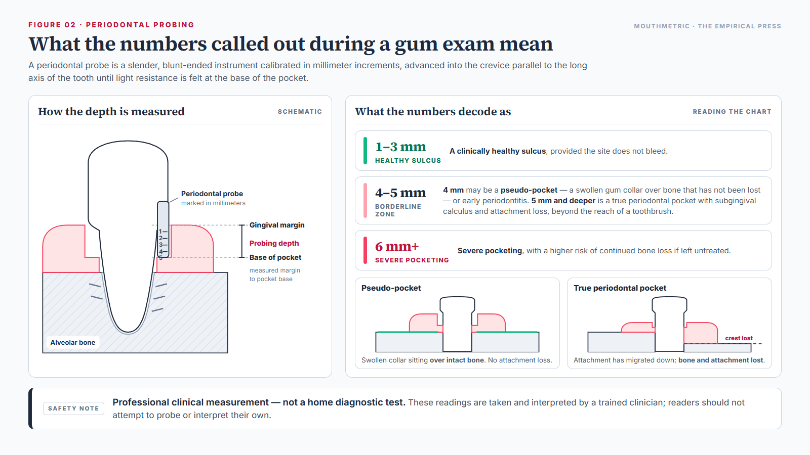
A periodontal probe is a slender, blunt-ended instrument calibrated in millimeter increments. The clinician advances it into the gum crevice parallel to the long axis of the tooth until light resistance is felt at the base of the pocket — roughly 0.20 to 0.25 Newtons of force. The numbers called out during an exam decode as follows:
- 1 to 3 mm: a clinically healthy sulcus, provided the site does not bleed.
- 4 mm: a borderline zone. It may be early periodontitis, or it may be a pseudo-pocket — a swollen gum collar that has grown upward with no bone loss at all.
- 5 mm and deeper: a true periodontal pocket, indicating subgingival calculus, attachment loss, and a root surface that a toothbrush cannot reach.
- 6 mm and deeper: severe pocketing, with a higher risk of continued bone loss if left untreated.
2. Clinical attachment level (CAL)
Probing depth alone does not measure true destruction. A tooth with severe recession can show a shallow pocket while substantial attachment has already been lost, and a tooth with drug-induced gum overgrowth can show a deep pocket with no bone loss at all. Clinicians therefore measure clinical attachment level (CAL): the distance in millimeters from a fixed landmark, the cementoenamel junction (CEJ) where enamel meets root cementum, to the base of the pocket. In practice, attachment level equals probing depth adjusted for where the gum margin sits relative to that landmark. CAL is the measure that defines periodontitis severity.
3. Bleeding on probing (BOP)
Whether capillary bleeding appears within 30 seconds of probing indicates active microscopic ulceration of the pocket lining. Under the 2017 World Workshop criteria, bleeding at fewer than 10% of sites is considered clinical gingival health, while bleeding at 30% or more of sites is classified as generalized gingivitis. Absence of bleeding is reassuring; it is not, on its own, proof of health, particularly in a smoker.
4. Full-mouth radiographs
Bitewing and periapical radiographs are needed to see what probing cannot. They show the level of the alveolar crest relative to the cementoenamel junction, whether bone loss is horizontal (uniform across adjacent teeth) or vertical and angular (trench-like defects along a root), and whether furcation involvement is present in multi-rooted teeth.
5. Periodontitis case definition
Under the international consensus published by Panos Papapanou and colleagues (2018), periodontitis is confirmed when interdental attachment loss is detectable at two or more non-adjacent teeth, or when attachment loss of 3 mm or more with pathological pockets deeper than 3 mm is detectable at two or more teeth, and the loss cannot be attributed to non-periodontal causes such as traumatic brushing abrasion, cervical caries, or endodontic lesions.
Understanding Periodontitis Stages and Grades (The 2017 AAP/EFP Framework)
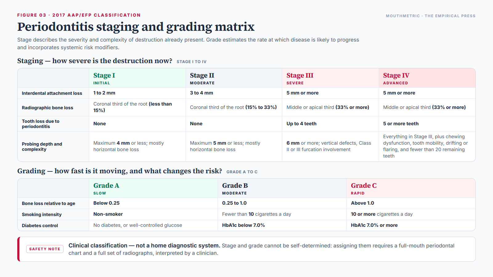
In 2017 the American Academy of Periodontology and the European Federation of Periodontology replaced the older “chronic” and “aggressive” labels with a framework modeled on oncology staging. Stage describes the severity and complexity of destruction already present. Grade estimates the rate at which disease is likely to progress and incorporates systemic risk modifiers. The point of the change was precision: two patients with the same depth of destruction can have entirely different trajectories.
The four stages
| Stage | Interdental attachment loss | Radiographic bone loss | Tooth loss to periodontitis | Probing depth and complexity |
|---|---|---|---|---|
| Stage I (initial) | 1 to 2 mm | Coronal third of the root (less than 15%) | None | Maximum probing depth of 4 mm or less; mostly horizontal bone loss |
| Stage II (moderate) | 3 to 4 mm | Coronal third of the root (15% to 33%) | None | Maximum probing depth of 5 mm or less; mostly horizontal bone loss |
| Stage III (severe, with potential for further tooth loss) | 5 mm or more | Extending into the middle or apical third of the root (33% or more) | Up to 4 teeth | Probing depth of 6 mm or more; vertical defects, Class II or III furcation involvement |
| Stage IV (advanced, with potential for loss of the dentition) | 5 mm or more | Extending into the middle or apical third of the root (33% or more) | 5 or more teeth | Everything in Stage III, plus chewing dysfunction, secondary bite trauma, tooth mobility, drifting or flaring of teeth, and fewer than 20 remaining teeth |
The three grades
Grading answers a different question from staging: not how bad it is now, but how fast it is moving. The primary grading criterion is bone loss relative to the patient’s age — how much attachment has been lost for how many years of life — supplemented by direct evidence of progression where a treatment history exists, and then by systemic modifiers:
- Grade A (slow): bone loss relative to age below 0.25; non-smoker; no diabetes or well-controlled glucose.
- Grade B (moderate): bone loss relative to age between 0.25 and 1.0; smoking fewer than 10 cigarettes a day; diabetes with HbA1c below 7.0%.
- Grade C (rapid): bone loss relative to age above 1.0; smoking 10 or more cigarettes a day; diabetes with HbA1c of 7.0% or more.
These stages are a clinical classification, not a home diagnostic system Stage and grade cannot be self-determined. Assigning them requires a full-mouth periodontal chart with measurements at every tooth and a full set of radiographs, interpreted by a clinician who can exclude non-periodontal causes of attachment loss. Nothing on this page, and no symptom you can observe yourself, is enough to place a mouth in a stage.
Staging and grading are not academic decoration. They set the treatment intensity, the re-evaluation interval, and the recall schedule that follows.
How Gum Disease Is Managed: The Clinical Treatment Pathway
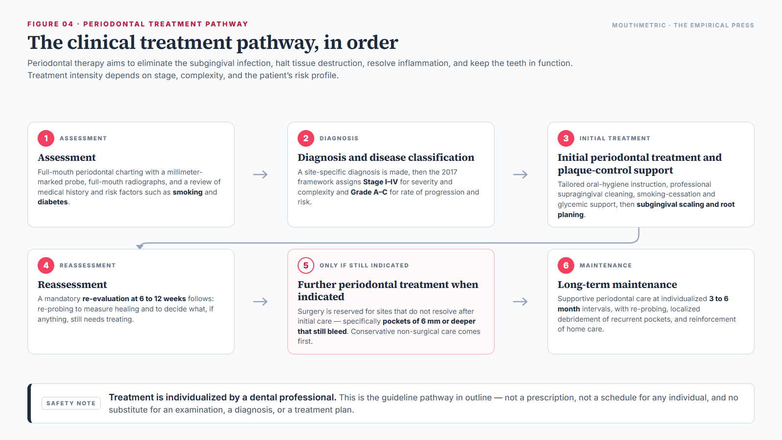
Periodontal therapy aims to eliminate the subgingival infection, halt tissue destruction, resolve inflammation, and keep the teeth in function. The sequence is codified in the European Federation of Periodontology’s S3-level clinical practice guideline (Sanz and colleagues, 2020) for Stages I to III, with a companion guideline for Stage IV (Herrera and colleagues, 2022). Treatment intensity depends on stage, complexity, and the patient’s risk profile, and it follows four ordered steps.
Step 1: Biofilm control and behavioural change
Therapy cannot work without the patient’s daily participation. Step 1 covers tailored oral hygiene instruction for brushing and interdental cleaning, professional supragingival cleaning to reduce the total microbial burden, smoking cessation support, and glycemic management for patients with diabetes.
Step 2: Subgingival instrumentation (scaling and root planing)
Scaling and root planing is the cornerstone of non-surgical periodontal therapy. Using ultrasonic instruments and hand curettes, the clinician removes subgingival calculus, biofilm, and bacterial endotoxins from root surfaces, usually under local anesthesia. In the American Dental Association’s systematic review and meta-analysis, Sharon Smiley and colleagues (2015) found that non-surgical instrumentation consistently achieves an average reduction in probing depth of 1 to 2 millimeters and an average gain in clinical attachment of 0.5 to 1.5 millimeters, with the largest gains in deeper pockets.
A mandatory re-evaluation at 6 to 12 weeks follows, to measure healing and decide what, if anything, still needs treating.
Step 3: Surgical therapy for pockets that persist
Surgery is reserved for sites that do not resolve after Steps 1 and 2 — specifically, pockets of 6 mm or deeper that still bleed. Options include access flap surgery for direct visualization and thorough debridement, resective surgery to reshape bone and reduce pocket depth, and regenerative surgery using bone grafts, barrier membranes, or enamel matrix derivative proteins in selected vertical defects. Regeneration is possible for specific infrabony defects; generalized horizontal bone loss cannot be regenerated, and it should not be presented as though it can.
Step 4: Supportive periodontal care
Periodontitis is a chronic condition, so treatment does not end. Supportive care means professional maintenance at individualized 3 to 6 month intervals, with re-probing, localized debridement of recurrent pockets, and reinforcement of home care. The value of that discipline was shown in the 30-year study by Per Axelsson, Jan Nyström, and Lars Lindhe: patients who maintained regular professional care over three decades lost an average of 0.4 to 0.7 teeth per person. Patients who stopped maintenance lost substantially more. Periodontitis can be arrested — but arrest is maintained, not achieved once.
Antibiotic stewardship Routine antibiotic pills are not recommended for periodontitis in Stages I to III, according to the EFP S3 guideline. Calcified subgingival biofilm has to be physically disrupted; a tablet cannot do that, and indiscriminate use drives antimicrobial resistance and gastrointestinal side effects. Adjunctive systemic antibiotics are reserved for specific Stage IV cases with rapid progression under the Stage IV guideline. Antibiotics are never a substitute for instrumentation, and no antibiotic should be taken for gum disease without a prescription and a diagnosis.
Can Gingivitis and Periodontitis Be Reversed?
The answer depends on which condition is present, and the honest version of the answer is more useful than a comforting one.
- Gingivitis: yes, completely. Because it is inflammation of soft tissue with no architectural breakdown, thorough plaque removal and professional cleaning resolve it — the tissues return to health within about a week to a fortnight of consistent care, with no permanent damage.
- Periodontitis: no, not the lost structure. Destroyed periodontal ligament fibers and resorbed alveolar bone do not regrow through brushing, flossing, mouthwash, supplements, or diet. No toothpaste rebuilds a bone crest.
- What is possible is arrest. Non-surgical therapy and supportive maintenance halt active destruction, stabilize the bone level, and allow the gum to reattach along a long junctional epithelium. The result is periodontal health on a reduced but stable foundation — teeth that are comfortable, functional, and retained.
On natural remedies: warm salt water soothes irritated tissue, and that is genuinely worth knowing when gums are sore. Essential oil rinses have modest adjunctive anti-plaque effects. But there is no high-quality trial evidence that oil pulling, salt rinses, herbal pastes, or supplements can halt periodontitis or rebuild bone, and none of them can mechanically remove calcified calculus from below the gumline. They are comforts, not treatments.
When to Seek Professional Care and Emergency Red Flags
Schedule a periodontal assessment if you notice any of the following:
- Gums that still bleed during brushing or flossing after 7 to 10 days of gentle, consistent care.
- Recurring swelling, soreness, or redness of the gum margins.
- Visible gum recession, or teeth that look longer than they used to.
- Chronic bad breath or a persistent bad taste that survives good oral hygiene.
- Any sensation of looseness, or a bite that has changed.
Red flags needing same-day or emergency care
Certain acute conditions do not wait for a routine appointment, as set out by David Herrera and colleagues (2018):
- Acute periodontal abscess: rapid-onset, throbbing, localized pain with swelling of the gum, pus draining from the margin, and pain on biting. Untreated, it can spread into the deep facial spaces.
- Necrotizing periodontal diseases (necrotizing ulcerative gingivitis and periodontitis): rapid destruction producing “punched-out” cratered gum papillae between teeth, intense burning pain, spontaneous bleeding, a gray pseudomembranous slough, fetid odor, and often swollen lymph nodes with fever.
- Systemic spread: any oral swelling accompanied by fever, swelling spreading under the jaw or toward the eye, or difficulty swallowing or breathing is a medical emergency requiring hospital assessment, because airway compromise is possible.
Evidence-Based Prevention and Daily Home Maintenance
Brushing
Brush twice daily for a full two minutes, angling the bristles about 45 degrees toward the gumline so the sulcular margin is cleaned — the modified Bass technique. Pressure should be light; aggressive scrubbing damages the gum margin it is meant to protect.
On equipment, the Cochrane review by Mohamed Yaacob and colleagues (2014) found that powered toothbrushes reduced plaque by 21% and gingival inflammation by 11% compared with manual brushes at three months, with oscillating-rotating heads giving the most consistent results. A manual brush still achieves excellent plaque control when technique is thorough, so the choice is preference and budget rather than a verdict on either.
Interdental cleaning
Bristles cannot reach between teeth. The Cochrane review by Helen Worthington and colleagues (2019) concluded that interdental brushes are significantly more effective than floss at removing interproximal plaque and reducing gum inflammation in open contacts. Floss remains the right tool where a brush cannot pass without forcing the tissue. Because the tongue carries a large share of oral biofilm, mechanical tongue scraping is a sensible addition — it reaches coating, though it cannot reach subgingival pockets.
Chemical plaque control as an adjunct
Antimicrobial rinses — chlorhexidine, cetylpyridinium chloride, and essential oil formulations — reduce plaque and inflammation when mechanical cleaning is compromised, for example after surgery or around a healing site. They are short-term adjuncts. They cannot penetrate a deep pocket, they do not remove calculus, and long-term unmonitored chlorhexidine use causes tooth staining and calculus buildup. For how rinses are evaluated by active ingredient, see our guide to antiseptic mouthwashes for bad breath. On clinical trials of oral probiotics, the evidence for adjunctive benefit remains early and should be described as such.
Daily practices beyond the mouth
Smoking cessation, glycemic control, and stress management all change the trajectory of gum disease. For the foundational daily routine these sit alongside, see our guide on how to get rid of bad breath, which covers brushing, interdental cleaning, and tongue hygiene in one sequence.
Common Misconceptions About Gum Disease
Myth 1: “Bacteria eat away at the jawbone.” Bacteria do not consume bone. Bone is resorbed by the body’s own osteoclasts, activated by inflammatory signaling — including the shift in the RANKL to OPG ratio described above. The damage is host-mediated, which is also why it does not stop on its own when the biofilm is left in place.
Myth 2: “If my gums don’t bleed, I don’t have gum disease.” Nicotine constricts the small vessels in the gum, so smokers frequently have pale, firm gums that do not bleed even with active, advanced disease. Bleeding is a useful signal in people who still produce it; its absence in a tobacco user is not evidence of health.
Myth 3: “A 4 mm pocket always means bone loss and a deep cleaning.” A 4 mm reading can be a pseudo-pocket — gum swelling that has raised the margin over intact bone. Swelling without attachment loss is gingivitis, and it resolves with plaque control. Determining which of the two a 4 mm reading represents requires probing, measurement of attachment level, and radiographs.
Myth 4: “Oil pulling or salt rinses can reverse gum disease.” Salt water soothes sore tissue, and that is its real benefit. No home remedy removes subgingival calculus, and none has trial evidence for regenerating lost bone. Mechanical debridement by a professional is what clears a true pocket.
Myth 5: “Gum disease causes heart attacks.” An association between periodontitis and cardiovascular disease exists and is documented. The American Heart Association’s review concluded that causation has not been demonstrated, and that treating gum disease has not been shown to prevent cardiac events. Neither the alarmist version nor the dismissive version of this claim is supported.
Myth 6: “Periodontitis inevitably means dentures.” It does not. In the 30-year Axelsson study, patients on regular maintenance lost an average of 0.4 to 0.7 teeth over three decades. The trajectory after diagnosis depends far more on treatment and maintenance than on the diagnosis itself.
Frequently Asked Questions
1. Is gingivitis the same thing as periodontitis?
No. Both fall under periodontal disease, but they represent different depths of damage. Gingivitis is mild, superficial inflammation of the gum tissue caused by plaque, with no loss of the bone or ligament fibers supporting the teeth, and it is fully reversible with thorough cleaning. Periodontitis is what happens when inflammation spreads into the supporting bone and periodontal ligament, causing permanent structural loss that cannot be undone at home.
2. Can gingivitis turn into periodontitis?
Periodontitis is always preceded by gingivitis, but not all gingivitis progresses. For inflammation to become destructive, several things have to align: persistent untreated subgingival biofilm, individual genetic or immune susceptibility, and modifying risk factors such as tobacco smoking, poorly controlled diabetes, chronic stress, and pregnancy-related hormonal change. Consistent daily plaque removal and regular professional cleanings are what keep gingivitis from ever crossing that line.
3. Can gum disease and bone loss be reversed at home?
Gingivitis can be fully reversed at home with diligent brushing, interdental cleaning, and professional cleanings. Once periodontitis has destroyed alveolar bone and ligament attachment, that structure does not regrow at home. No special toothpaste, mouthwash, oil pulling routine, or supplement rebuilds lost jawbone. What professional treatment combined with excellent home care can do is halt active destruction, stabilize the remaining bone, and prevent further tooth loss.
4. Can you have severe gum disease without bleeding or pain?
Yes. Chronic periodontitis is often called a silent disease because it rarely causes pain in its early and moderate stages. Smoking makes this worse: nicotine-induced vasoconstriction narrows the vessels in the gums and markedly suppresses bleeding, so a heavy smoker can have pale, firm gums that never bleed while bone loss continues underneath. This is why periodic professional probing and dental x-rays matter even when the mouth feels completely normal.
5. What do the pocket depth numbers called out during a gum exam mean?
A clinician uses a millimeter-marked probe to measure the space between tooth and gum. 1 to 3 mm indicates a healthy crevice. 4 mm is a borderline zone that may be early periodontitis or a swollen pseudo-pocket with no bone loss. 5 mm or deeper indicates a true periodontal pocket where bacteria and calculus have accumulated beyond the reach of a toothbrush, typically calling for professional deep cleaning. 6 mm and deeper is severe pocketing. These are clinical measurements performed and interpreted by a trained professional; they are not readings a patient can take or interpret at home.
6. Why did my dentist recommend a “deep cleaning” instead of a regular cleaning?
A standard cleaning (prophylaxis) removes plaque and tartar from tooth surfaces above the gumline in a healthy mouth. When pockets reach 5 mm or deeper with bone loss, bacteria and hardened calculus sit on the root surfaces below the gumline where a regular cleaning cannot reach. Scaling and root planing is the therapeutic procedure that addresses that: performed under local anesthesia, it physically debrides root surfaces, removes bacterial endotoxins, and allows the gum to heal and tighten. On average it reduces pocket depth by 1 to 2 mm and gains 0.5 to 1.5 mm of attachment.
7. Does gum disease cause bad breath?
Yes, through a specific mechanism. Deep pockets create an oxygen-poor environment for anaerobic bacteria such as Porphyromonas gingivalis and Treponema denticola. These break down proteins in blood and gingival crevicular fluid, releasing volatile sulfur compounds — notably methyl mercaptan (CH₃SH). Because these organisms live below the gumline inside pockets, mints, mouthwashes, and tongue scrapers cannot reach them; only professional subgingival debridement removes the source.
8. When does gum swelling or bleeding become a dental emergency?
Routine bleeding during brushing warrants a dental check-up but is not an emergency. Seek same-day care for severe throbbing pain with localized gum swelling and pus drainage, which suggests a periodontal abscess; for rapid, severe pain with gray, bleeding, “punched-out” tissue between the teeth, which suggests necrotizing disease; or for any gum swelling accompanied by fever, facial swelling, or difficulty swallowing or breathing, which can indicate a spreading infection needing hospital assessment.
Bottom Line
Gum disease is a chronic inflammatory spectrum: superficial, fully reversible gingivitis at one end, and destructive periodontitis — with permanent loss of ligament and bone — at the other. Gingivitis does not automatically become periodontitis, but progression is painless and invisible, so the deciding factor is usually how early it is found rather than how much it hurts.
Because periodontitis advances silently, regular professional examinations, full-mouth probing, and radiographs are what establish where you actually stand. Gingivitis resolves with professional care and daily plaque control. Established periodontitis is arrested rather than reversed, and keeping it arrested depends on treatment followed by maintenance at 3 to 6 month intervals.
Sources
- Chapple ILC, Mealey BL, Van Dyke TE, et al. (2018). Periodontal health and gingival diseases and conditions on an intact and a reduced periodontium: consensus report of workgroup 1 of the 2017 World Workshop on the Classification of Periodontal and Peri-Implant Diseases and Conditions. Journal of Periodontology, 89 Suppl 1, S74–S84. PMID 29926944. https://pubmed.ncbi.nlm.nih.gov/29926944/
- Caton JG, Armitage G, Berglundh T, et al. (2018). A new classification scheme for periodontal and peri-implant diseases and conditions — introduction and key changes from the 1999 classification. Journal of Clinical Periodontology, 45 Suppl 20, S1–S8. PMID 29926489. https://pubmed.ncbi.nlm.nih.gov/29926489/
- Löe H, Theilade E, Jensen SB (1965). Experimental gingivitis in man. Journal of Periodontology, 36(3), 177–187. PMID 14296927. https://pubmed.ncbi.nlm.nih.gov/14296927/
- Eke PI, Dye BA, Wei L, et al. (2012). Prevalence of periodontitis in adults in the United States: 2009 and 2010. Journal of Dental Research, 91(10), 914–920. PMID 22935673. https://pubmed.ncbi.nlm.nih.gov/22935673/
- Eke PI, Dye BA, Wei L, et al. (2015). Update on prevalence of periodontitis in adults in the United States: NHANES 2009 to 2012. Journal of Periodontology, 86(5), 611–622. PMID 25688694. https://pubmed.ncbi.nlm.nih.gov/25688694/
- Eke PI, Thornton-Evans GO, Wei L, et al. (2018). Periodontitis in US adults: National Health and Nutrition Examination Survey 2009–2014. Journal of the American Dental Association, 149(7), 576–588.e6. PMID 29957185. https://pubmed.ncbi.nlm.nih.gov/29957185/
- Hajishengallis G, Darveau RP, Curtis MA (2012). The keystone-pathogen hypothesis. Nature Reviews Microbiology, 10(10), 717–725. PMID 22941505. https://pubmed.ncbi.nlm.nih.gov/22941505/
- Socransky SS, Haffajee AD, Cugini MA, et al. (1998). Microbial complexes in subgingival plaque. Journal of Clinical Periodontology, 25(2), 134–144. PMID 9495612. https://pubmed.ncbi.nlm.nih.gov/9495612/
- Sorsa T, Gursoy UK, Nwhator S, et al. (2016). Analysis of matrix metalloproteinases, especially MMP-8, in gingival crevicular fluid, mouthrinse and saliva for monitoring periodontal diseases. Periodontology 2000, 70(1), 142–163. PMID 26662488. https://pubmed.ncbi.nlm.nih.gov/26662488/
- Belibasakis GN, Bostanci N (2012). The RANKL–OPG system in clinical periodontology. Journal of Clinical Periodontology, 39(3), 239–248. PMID 22092994. https://pubmed.ncbi.nlm.nih.gov/22092994/
- Yaegaki K, Sanada K (1992). Volatile sulfur compounds in mouth air from clinically healthy subjects and patients with periodontal disease. Journal of Periodontal Research, 27(4 Pt 1), 233–238. PMID 1640345. https://pubmed.ncbi.nlm.nih.gov/1640345/
- Tomar SL, Asma S (2000). Smoking-attributable periodontitis in the United States: findings from NHANES III. Journal of Periodontology, 71(5), 743–751. PMID 10872955. https://pubmed.ncbi.nlm.nih.gov/10872955/
- Dietrich T, Bernimoulin JP, Glynn RJ (2004). The effect of cigarette smoking on gingival bleeding. Journal of Periodontology, 75(1), 16–22. PMID 15025212. https://pubmed.ncbi.nlm.nih.gov/15025212/
- Preshaw PM, Alba AL, Herrera D, et al. (2012). Periodontitis and diabetes: a two-way relationship. Diabetologia, 55(1), 21–31. PMID 22057194. https://pubmed.ncbi.nlm.nih.gov/22057194/
- Michalowicz BS, Diehl SR, Gunsolley JC, et al. (2000). Evidence of a substantial genetic basis for risk of adult periodontitis. Journal of Periodontology, 71(11), 1699–1707. PMID 11128917. https://pubmed.ncbi.nlm.nih.gov/11128917/
- Chen P, Hong F, Yu X (2022). Prevalence of periodontal disease in pregnancy: a systematic review and meta-analysis. Journal of Dentistry, 125, 104253. PMID 35998741. https://pubmed.ncbi.nlm.nih.gov/35998741/
- Bobetsis YA, Graziani F, Gürsoy M, Madianos PN (2020). Periodontal disease and adverse pregnancy outcomes. Periodontology 2000, 83(1), 154–174. PMID 32385871. https://pubmed.ncbi.nlm.nih.gov/32385871/
- Seymour RA, Ellis JS, Thomason JM (2000). Risk factors for drug-induced gingival overgrowth. Journal of Clinical Periodontology, 27(4), 217–223. PMID 10783833. https://pubmed.ncbi.nlm.nih.gov/10783833/
- Lockhart PB, Bolger AF, Papapanou PN, et al. (2012). Periodontal disease and atherosclerotic vascular disease: does the evidence support an independent association? A scientific statement from the American Heart Association. Circulation, 125(20), 2520–2544. PMID 22514251. https://pubmed.ncbi.nlm.nih.gov/22514251/
- Tonetti MS, Greenwell H, Kornman KS (2018). Staging and grading of periodontitis: framework and proposal of a new classification and case definition. Journal of Periodontology, 89 Suppl 1, S159–S172. PMID 29926952. https://pubmed.ncbi.nlm.nih.gov/29926952/
- Papapanou PN, Sanz M, Buduneli N, et al. (2018). Periodontitis: consensus report of workgroup 2 of the 2017 World Workshop on the Classification of Periodontal and Peri-Implant Diseases and Conditions. Journal of Periodontology, 89 Suppl 1, S173–S182. PMID 29926951. https://pubmed.ncbi.nlm.nih.gov/29926951/
- Sanz M, Herrera D, Kebschull M, et al. (2020). Treatment of stage I–III periodontitis — the EFP S3 level clinical practice guideline. Journal of Clinical Periodontology, 47 Suppl 22, S4–S60. PMID 32383274. https://pubmed.ncbi.nlm.nih.gov/32383274/
- Herrera D, Sanz M, Kebschull M, et al. (2022). Treatment of stage IV periodontitis: the EFP S3 level clinical practice guideline. Journal of Clinical Periodontology, 49 Suppl 24, S4–S71. PMID 35688447. https://pubmed.ncbi.nlm.nih.gov/35688447/
- Smiley CJ, Tracy SL, Abt E, et al. (2015). Systematic review and meta-analysis on the nonsurgical treatment of chronic periodontitis by means of scaling and root planing with or without adjuncts. Journal of the American Dental Association, 146(7), 508–524.e5. PMID 26113099. https://pubmed.ncbi.nlm.nih.gov/26113099/
- Herrera D, Retamal-Valdes B, Alonso B, Feres M (2018). Acute periodontal lesions (periodontal abscesses and necrotizing periodontal diseases) and endo-periodontal lesions. Journal of Periodontology, 89 Suppl 1, S85–S102. PMID 29926942. https://pubmed.ncbi.nlm.nih.gov/29926942/
- Axelsson P, Nyström B, Lindhe J (2004). The long-term effect of a plaque control program on tooth mortality, caries and periodontal disease in adults. Results after 30 years of maintenance. Journal of Clinical Periodontology, 31(9), 749–757. PMID 15312097. https://pubmed.ncbi.nlm.nih.gov/15312097/
- Worthington HV, MacDonald L, Poklepovic Pericic T, et al. (2019). Home use of interdental cleaning devices, in addition to toothbrushing, for preventing and controlling periodontal diseases and dental caries. Cochrane Database of Systematic Reviews, 4(4), CD012018. PMID 30968949. https://pubmed.ncbi.nlm.nih.gov/30968949/
- Yaacob M, Worthington HV, Deacon SA, et al. (2014). Powered versus manual toothbrushing for oral health. Cochrane Database of Systematic Reviews, (6), CD002281. PMID 24934383. https://pubmed.ncbi.nlm.nih.gov/24934383/