Learn
Receding Gums: Causes, Measurement, Treatment, and What to Know
What are receding gums? Causes, how dentists measure recession, RT1–RT3, when monitoring is enough, and how predictable root coverage really is.
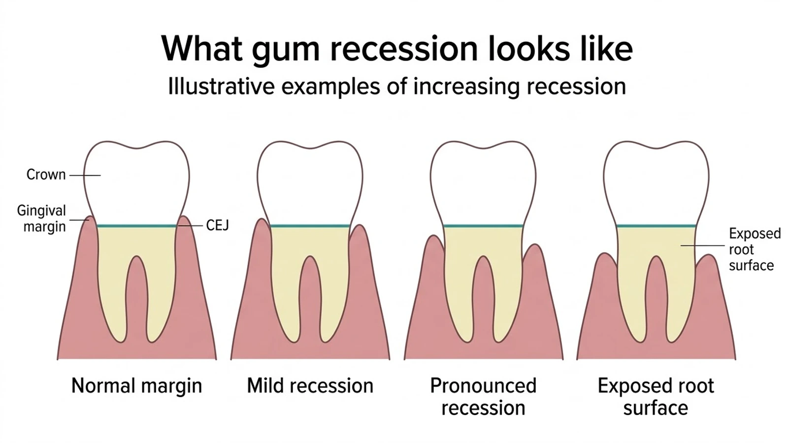
If you have noticed that one tooth looks a little longer than it used to — or that the gumline around it sits lower than its neighbors, leaving a yellower surface visible above the tooth — you are looking at what clinicians call gingival recession, or receding gums. It is one of the most common things people notice about their own mouths, and one of the least well explained on the internet.
Recession is not a disease in itself. It is a finding: a measurable change in where the gum margin sits relative to the tooth. Behind that finding sits a long list of possible contributors — anatomy, inflammation, technique, habit, and time — and a clinical decision that is more nuanced than “you need surgery” or “you’re brushing too hard.” This article explains what recession actually is, how clinicians measure and classify it, what the evidence does and does not say about its causes, and when monitoring is a perfectly reasonable plan.
MouthMetric Medical Evidence Review
Every clinical, numeric, and classification claim in this article is traced to a cited peer-reviewed study, systematic review, or consensus document, with full references at the end of the page. We do not diagnose, and this page cannot assess your individual situation: recession depth, attachment level, and bone support can only be measured by a licensed dental professional using a calibrated probe and radiographs. Nothing here replaces an examination, a diagnosis, or a treatment plan, and no passage should be read as a reason to delay care or to treat yourself at home. For how we select, grade, and update evidence, see the MouthMetric evidence methodology.
What Are Receding Gums?
Gum recession is the apical migration of the gingival margin — the gumline has moved down the root of the tooth, away from the chewing surface. What makes that movement definable rather than subjective is a fixed landmark called the cementoenamel junction (CEJ): the line where the enamel of the crown meets the cementum covering the root. In health, the gum margin sits at or slightly above that line. When the margin sits apical to it — lower, toward the root tip — the root surface is exposed, and the tooth can appear longer than its neighbors.
Three points keep the definition honest:
- Recession exposes root surface, not just “gaps.” The tissue that was covering the root is gone from that spot, which is why exposed areas may look yellower than enamel, feel different to a soft brush, and respond differently to temperature.
- Gum margin position varies naturally between teeth and between people. Some mouths have scalloped, thick margins in some areas and thinner, more apical margins in others — particularly on the outer (buccal) surfaces, where recession is most commonly found. Change over time matters more than a single photograph.
- Recession is a sign, not a diagnosis. A dentist records it, measures it, and interprets it alongside everything else in the mouth. It does not, on its own, name a condition — and it is emphatically not something to classify from a mirror.
How Common Is Gum Recession?
Very common — but “common” depends entirely on how the question is asked, and different well-conducted studies give different-looking numbers for reasons that are mostly about thresholds and methods rather than disagreement.
The most recent pooled estimate comes from a 2025 systematic review and meta-analysis of 22 observational studies. Depending on how deep you set the bar, the authors estimated:
| Threshold | Pooled prevalence (95% CI) |
|---|---|
| ≥ 1 mm recession at one or more sites | 81.1% (73.9–86.7) |
| ≥ 3 mm recession at one or more sites | 48.4% (39.7–57.2) |
| ≥ 5 mm recession at one or more sites | 16.2% (9.1–27.4) |
Source: Marschner and colleagues, 2025.
The national US survey data tell a compatible story at a different threshold. In NHANES III — 9,689 US adults aged 30 and older, examined between 1988 and 1994 — Albandar and Kingman estimated that 22.5% of US adults (about 23.8 million people) had at least one tooth surface with 3 mm or more of recession, with prevalence and severity rising steadily with age.
Earlier reviews reach the same shape of conclusion from different surveys: Kassab and Cohen, reviewing the epidemiologic literature in the Journal of the American Dental Association, reported surveys in which 88% of people aged 65 and older and about 50% of people aged 18 to 64 had one or more sites with recession. A separate global systematic review covering studies published from 1991 to 2021 (Yadav and colleagues, 2023) likewise concluded that recession is highly prevalent across populations.
The age gradient has a forward edge, too. A critical review in the Journal of Clinical Periodontology (Heasman and colleagues, 2017) evaluated treatment evidence for gingival recession and root caries together and concluded that both conditions will become more prevalent as patients retain their teeth for longer — recession and its consequences scale with how long people keep their teeth, not just with how old they are.
Why the numbers look so different across studies — 81% versus 22% — is worth understanding rather than glossing over:
- Threshold. Any visible 1 mm defect is counted in one figure; only 3 mm defects are counted in another.
- Sites examined. Some surveys measure two surfaces in two quadrants; others chart every surface.
- Age and population. Recession increases with age; a study of older adults will report more than one of young adults.
- Method. Probe technique, calibration, and whether interproximal surfaces are included all move the numbers.
None of these figures describes you, and none of them is a prediction. What they establish is that recession is an ordinary feature of adult mouths — common enough that finding some is not, by itself, alarming, and common enough that it deserves a proper explanation rather than a scare headline.
What Causes Gum Recession?
Recession is multifactorial. Almost every page you will read online reduces it to one sentence — “brushing too hard causes receding gums” — and the evidence does not support that reduction. What follows is the causal picture as the current evidence actually describes it.
Anatomy: the tissue you started with
The 2018 World Workshop review of mucogingival conditions (Cortellini and Bissada) concluded that thin periodontal phenotypes — the clinical term for the delicate, scalloped gum type, often still called a “biotype” — are at greater risk of developing recession, and that recession increases with age and occurs in populations with both high and low standards of oral hygiene. Local anatomy matters too: in the 2025 meta-analysis, a high frenulum (a muscle attachment that pulls on the gum margin) was associated with recession at an odds ratio of 4.58. Anatomical risk is not a fault; it is a starting condition that changes how much small insults matter.
Periodontal inflammation
The strongest statistical association in the 2025 meta-analysis was periodontitis itself (OR 9.90, 95% CI 4.15–23.60), with dental plaque (OR 4.26, 95% CI 2.91–6.24) also strongly associated. This is the main bridge to the separate topic of gum disease: causes, stages, and clinical management — when bone and ligament support are lost, the gum margin follows them down. Inflammation and recession frequently coexist; neither one explains the other completely.
Toothbrushing: the honest version
This is where most health pages oversimplify, so it is worth being precise about what the studies found.
A dedicated systematic review by Heasman and colleagues (2015) set out to answer exactly this question and concluded that “the data to support or refute the association between toothbrushing and gingival recession and non-carious cervical lesions remain largely inconclusive.” Their meta-analysis covered just 159 subjects; thirteen cross-sectional studies identified the brushing factors most consistently associated with recession — scrub or horizontal technique, higher frequency, longer duration, harder bristles, and infrequent brush replacement — but cross-sectional associations cannot establish cause.
Two controlled trials point the same cautious direction:
- In a 12-month randomized trial (McCracken and colleagues, 2009), there was no progression of gingival recession in subjects using either a manual or a powered toothbrush.
- In a three-year randomized study (Dörfer and colleagues, 2016), pre-existing recession in healthy adults decreased with both brush types — from 2.35 mm to 1.90 mm with the power brush and from 2.26 mm to 1.81 mm with the manual brush, with no difference between groups. (The study was funded by a manufacturer; the authors suggested that participants’ heightened awareness and gentler technique over three years may explain the reduction.)
The fair reading: aggressive, high-pressure, horizontal scrubbing of the margin is a plausible contributor in some people — clinicians see it, and cross-sectional data keep pointing at it — but the population-level evidence remains mixed, and proper brushing does not, on average, make recession worse. Technique is worth correcting; panic and under-brushing are not the answer.
Tooth position, orthodontics, and restorations
The same World Workshop review found that inadequate oral hygiene, orthodontic treatment, and cervical restorations (fillings at the gumline) might increase the risk of developing recession. Teeth that sit prominently in the arch — pushed outward in bone that does not fully cover the root — are a classic site for recession, which is also why the buccal surfaces of upper front teeth appear so often in clinical series.
Smoking, alcohol, and other associated factors
From the same 2025 meta-analysis of observational studies, smoking, alcohol consumption, male sex, occlusal trauma, and a history of periodontal treatment were each significantly associated with recession; the full odds ratios and confidence intervals sit in the table below. Two cautions belong with those numbers. First, the intervals are wide for the strongest estimates — the periodontitis figure, for instance, ranges from about 4 to about 24 — so the precise magnitude stays uncertain even where the direction is consistent. Second, not every study agrees: a study of smokers with minimal periodontal disease (Müller and colleagues, 2002) did not find that smokers were at increased risk of developing recession. These are associations drawn from observational data, not proven causes, and the honest way to present them is as factors your clinician will weigh rather than verdicts.
| Factor (2025 meta-analysis, 22 studies) | Odds ratio (95% CI) |
|---|---|
| Periodontitis | 9.90 (4.15–23.60) |
| High frenulum | 4.58 (2.58–8.11) |
| Dental plaque | 4.26 (2.91–6.24) |
| Occlusal trauma | 3.20 (1.74–5.87) |
| Alcohol consumption | 2.04 (1.51–2.75) |
| History of periodontal treatment | 1.86 (1.33–2.58) |
| Smoking | 1.84 (1.33–2.53) |
| Male sex | 1.52 (1.36–1.69) |
Odds ratios from meta-analyses of observational studies. An odds ratio above 1 indicates association, not causation.
Taken together, the causes section of most consumer pages is a single sentence too thin. The real picture: an anatomical starting point, an inflammatory driver, a set of local mechanical factors, habit and behavior, and time.
Does Gum Recession Mean You Have Gum Disease?
No — not by itself, and this distinction matters for how alarmed you should feel.
Recession occurs in populations with both high and low standards of oral hygiene (Cortellini and Bissada, 2018), and it has been found in patients with good and poor hygiene alike (Kassab and Cohen, 2003). Plenty of people have localized recession with healthy pockets, no bleeding, intact interproximal attachment, and no periodontitis at all.
At the same time, the relationship is not zero. Periodontitis was the strongest factor associated with recession in the 2025 meta-analysis, because when supporting bone and ligament are destroyed, the gum margin — which has nothing left to sit on — moves with them. And because recession changes the math of a periodontal exam (more on that below), the two findings are often recorded together.
The practical implications:
- Recession alone does not diagnose periodontitis. A periodontal diagnosis requires probing of pocket depths and attachment levels at multiple sites, assessment of bleeding, and radiographs — a structured examination, not a look in the mirror. Our companion article covers gum disease causes, stages, and clinical management in full, including how staging and grading work.
- Periodontitis alone does not prove recession caused it, or vice versa. They can travel together; they are not the same condition.
- Bleeding and inflammation change the picture. Recession on a firm, non-bleeding margin in a stable mouth is a different clinical situation from recession alongside bleeding, deep pockets, or bone loss — which is one reason a professional assessment considers the broader periodontal picture rather than the single most visible finding.
How Do Dentists Measure Gum Recession?
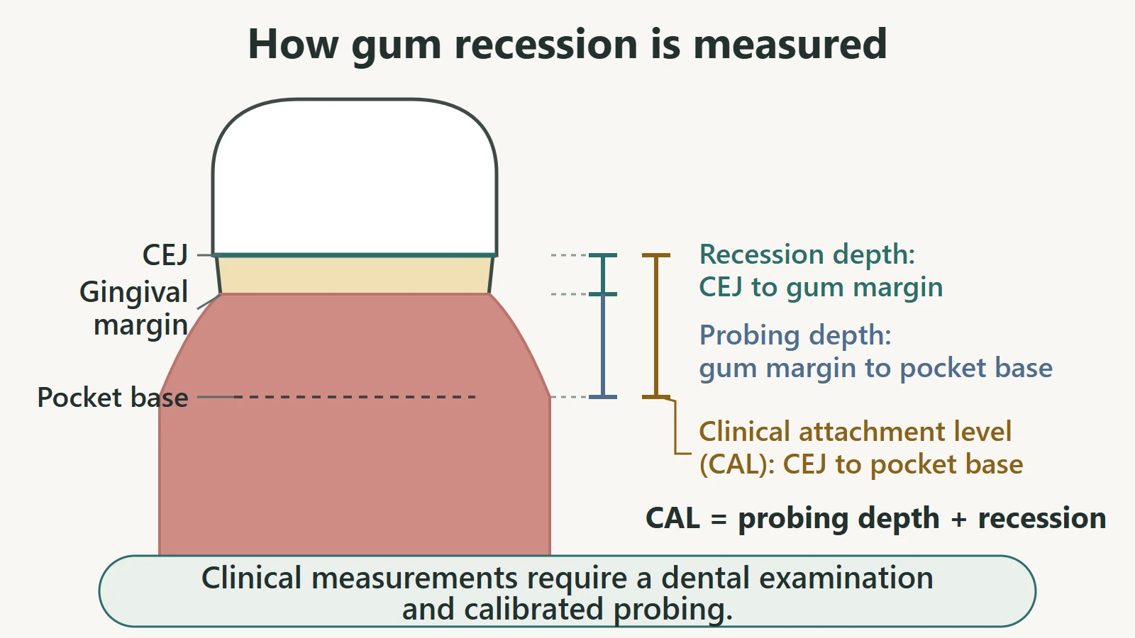
Measurement is what turns “my gum looks lower” into a clinical finding. Three distances, all anchored to the same fixed landmark, do the work:
| Measurement | From | To | What it shows |
|---|---|---|---|
| Gingival recession | CEJ (enamel–cementum line) | Gum margin | How far the margin has migrated apically, in millimeters |
| Probing depth | Gum margin | Base of the crevice/pocket | How deep the space is right now |
| Clinical attachment level (CAL) | CEJ | Base of the crevice/pocket | Total attachment lost at that site |
The arithmetic is the point. Clinical attachment level equals probing depth plus recession when the margin sits apical to the CEJ. That is why a tooth can show a perfectly shallow pocket reading and still have lost substantial attachment: the measuring point (the margin) has moved. It is the same principle our gum disease article uses to explain why clinicians chart CAL and not pocket depth alone — a tooth with severe recession can show a shallow pocket while substantial attachment has already been lost.
In practice, a clinician records these values at multiple surfaces around each tooth, because recession is frequently a localized, surface-specific finding — most often on the buccal aspect — while interproximal attachment tells a separate part of the story (which is exactly what the classification in the next section uses).
This is a clinical procedure, not a home measurement These values are taken with a calibrated millimeter probe under controlled conditions and interpreted with radiographs. A mirror, a photograph, or a ruler held against the tooth cannot give you a meaningful recession measurement or an attachment level, and no reading taken at home should be used to make treatment decisions. If you want to know where you stand, that is a question for a chair, not a screen.
What Are RT1, RT2, and RT3?
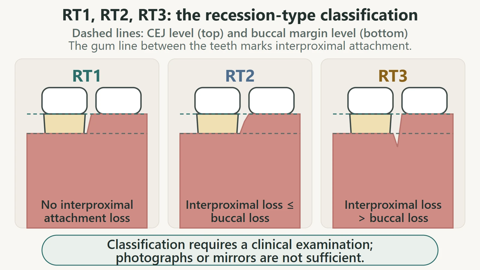
Because the shape of a recession defect predicts what treatment can achieve, clinicians classify them. The system now used most widely in periodontal literature was proposed by Cairo and colleagues in 2011, and it turns on a single question: how much attachment loss is there between the teeth, compared with the visible defect on the outer surface?
| Type | Interproximal attachment (between the teeth) | What it signals about prognosis |
|---|---|---|
| RT1 | No interproximal attachment loss | The conditions for complete root coverage are most favorable |
| RT2 | Interproximal loss present, but less than or equal to the loss at the buccal site | Complete coverage may be achievable, but interproximal loss limits reduction |
| RT3 | Interproximal loss greater than at the buccal site | Full root coverage is not achievable; goals are set accordingly |
In the original validation study — 116 recession defects in 25 patients, average defect depth 3.2 mm — the classification was reliable between examiners (inter-rater correlation 0.86) and predicted the final reduction in recession at six months (p < 0.0001). Its value is exactly that: it converts a visible defect into an honest prognosis before anyone operates.
A classification is not a self-diagnosis tool RT categories depend on measured interproximal attachment — values taken with a probe between the teeth, beside the defect — not on appearance. You cannot assign yourself a type from a photograph, a mirror, or a ring light, and this page does not tell you how to. The categories exist so that a clinician can describe a defect and discuss realistic goals with you; if someone (including a website) tells you your type based on a picture, treat that as a marketing claim, not an assessment.
Do Receding Gums Grow Back?
The direct answer: receding gum tissue does not spontaneously migrate back to its original position. The margin does not regenerate to where it used to be on its own, and the evidence reviewed for this article contains no toothpaste, supplement, rinse, or device shown to put it back.
Three qualifications keep that answer honest rather than bleak:
- Measurements can change without true regeneration. In the three-year brushing trial described above, recorded recession depth decreased in both groups — and the authors attributed that at least partly to participants brushing more gently as the study progressed. Tissue that is inflamed, swollen, or traumatized can settle, and a margin can read slightly differently as the local conditions change. Improvement in a measurement is not the same thing as tissue regenerating to its former position.
- Recession can also move the other way. A systematic review of untreated defects (Chambrone and Tatakis, 2016) followed 1,647 recession defects across six studies for at least 24 months: 78.1% of defects deepened over the follow-up period, and pooled estimates showed significantly increased odds of developing new recession over time — even in individuals with good oral hygiene. Not every defect progresses, and not at the same rate, but “leave it alone and it will reverse” is not what the evidence shows.
- Professional treatment can improve coverage — within limits. Root-coverage procedures can partially or sometimes fully cover an exposed root in suitable cases. What they cannot do is regenerate a ligament-and-bone complex or guarantee a return to where the tissue once sat. Expectations are set by the defect (see the RT classification above), the tissue you started with, and the numbers in the next-but-one section.
When Can Gum Recession Be Monitored?
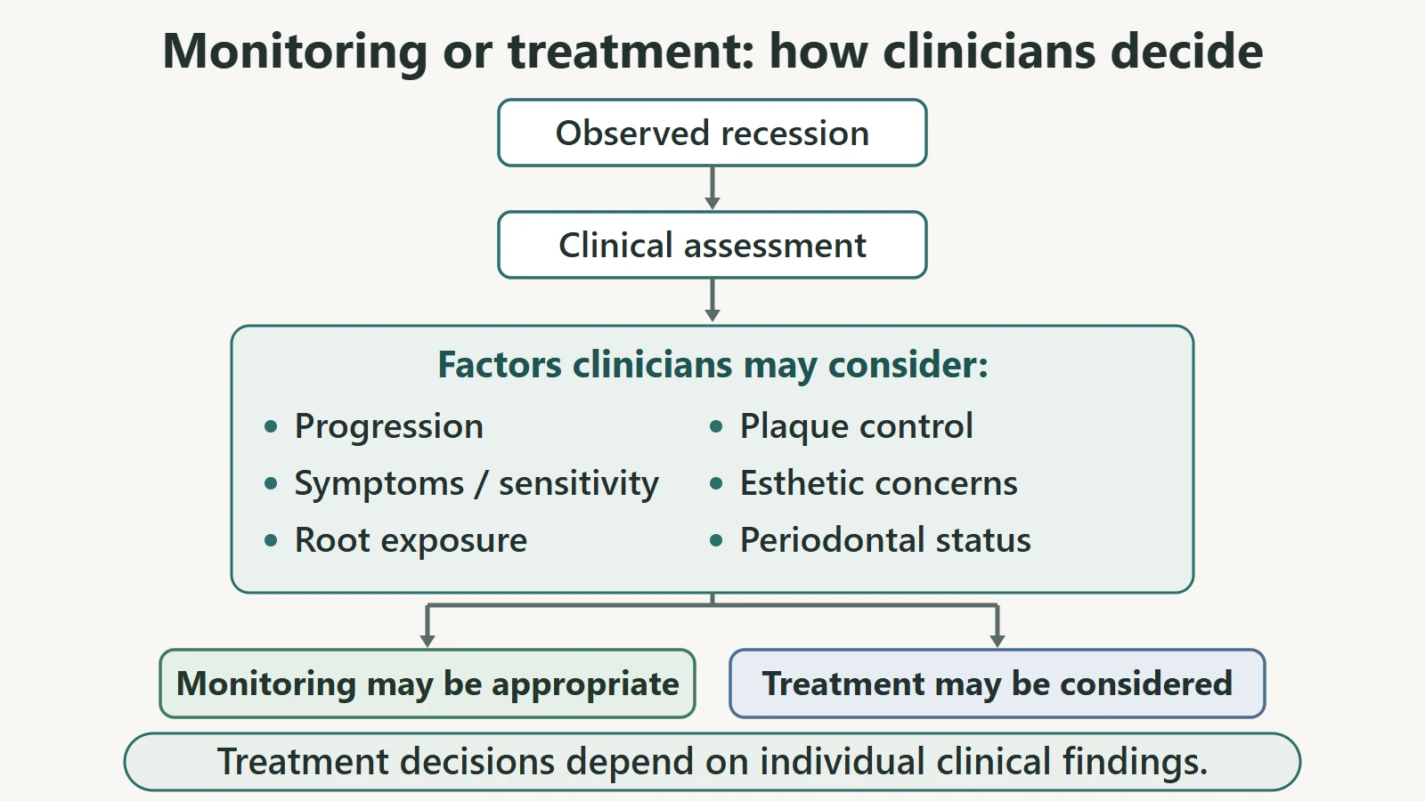
Not every recession defect requires surgery, and the consensus review that frames modern mucogingival care says so explicitly. The 2018 World Workshop review (Cortellini and Bissada) concluded that “in the absence of pathosis, monitoring specific sites seems to be the proper approach” — and that periodontal health can be maintained in most patients with optimal home care.
Monitoring is a clinical decision, made with you, based on factors such as:
- Progression — whether measurements are stable over successive visits or deepening.
- Symptoms — whether the site is sensitive, tender, or causing trouble.
- Root exposure — how much root surface is exposed and where.
- Root-surface health — whether the exposed surface is developing cavities or non-carious cervical lesions (the small notches that form at the gumline). Root exposure is frequently associated with these changes, along with dentin hypersensitivity and esthetic concerns.
- Plaque control — whether the site can be kept clean comfortably and consistently.
- Periodontal status — pockets, bleeding, attachment, and bone levels elsewhere in the mouth.
- Phenotype and local factors — tissue thickness, tooth position, restoration margins, and orthodontic context.
- Your own priorities — the esthetic importance of the site to you is a legitimate input; the consensus review lists patient esthetic requirements as one of the reasons intervention may be indicated.
None of these factors can be weighed properly from outside a clinical relationship, and monitoring is an active plan — periodic re-measurement, not neglect. This section describes how clinicians think; it does not tell you which category you are in.
When Is Treatment Considered?
Guidance from the same consensus review frames the indications generally: surgical intervention — either to change the tissue phenotype or to cover the root — may be considered when the risk of developing or progressing damage is increased, or to satisfy a patient’s esthetic requirements.
In practice, the reasons clinicians raise treatment tend to include:
- Progressive recession — documented deepening between visits despite good home care.
- Persistent or troublesome sensitivity that affects normal eating or brushing and does not settle.
- Ongoing root exposure of concern, particularly where the exposed surface is at risk of cavities or cervical lesions.
- Difficulty keeping the site clean — plaque control compromised by the anatomy of the defect.
- Significant esthetic concern, particularly on visible upper front teeth.
- Combined periodontal findings, where recession is part of a wider picture being managed.
Which of these applies — and which procedure, if any, fits — is a decision made after examination, measurement, and a conversation about goals. This page deliberately does not tell you which procedure you personally need; that question can only be answered by someone who has seen the site.
How Predictable Is Gum-Recession Treatment?
This is where realistic expectations are won or lost, so the numbers deserve their full context.
The reference procedure. The most extensively studied approach combines a subepithelial connective tissue graft (tissue taken from the palate) with a coronally advanced flap (the gum repositioned over the root). An updated Cochrane review of 48 randomized trials (Chambrone and colleagues, 2019) found greater recession reduction with this combination than with guided tissue regeneration plus flap, and greater gains in keratinized tissue width than several alternatives. It also found insufficient evidence of a difference between the graft and two common substitute materials in some comparisons — an honest “we don’t know yet,” not an endorsement of either. A systematic review for the AAP Regeneration Workshop (Chambrone and Tatakis, 2015) reached the same field-level picture: these procedures work, with technique and defect selection determining the result.
The long-term numbers — with their boundaries attached. One 20-year follow-up, by Pini Prato and colleagues (2018), tracked a private-practice cohort of 45 patients with 45 single recession defects on upper teeth, treated between 1990 and 1997. Mean root coverage was 74.23% at one year and 67.69% at 20 years — that is, most of the gain held, with some gradual relapse. Complete coverage occurred in about 57% of the simpler (Class I) defects at one year, falling to about 48% at 20 years. Recurrence was associated with less than 2 mm of attached keratinized tissue, the presence of root steps, and smoking.
Read those figures for what they are: one cohort, 45 upper-tooth defects, treated by the technique of that era, classified by the older Miller system. They are not a universal success rate, not a guarantee, and not applicable to every defect or every patient. Two defects in two mouths can behave differently — which is precisely why classification, phenotype, and history are assessed before anyone quotes a number at you.
Newer techniques promoted directly to consumers — including approaches that avoid a palatal donor site — have a substantially shorter evidence history than graft-plus-flap. A registered randomized comparison of one such technique against connective tissue graft enrolled 43 participants and completed follow-up in 2023 (ClinicalTrials.gov NCT04356391) — a single small trial, measured in years, set against more than three decades of graft-plus-flap data. A young evidence base is a reason to ask questions, not a reason to assume either that a technique works or that it does not.
The overall picture: root-coverage procedures are legitimate, well-studied interventions with good short-term results that soften somewhat over decades, and with outcomes that depend heavily on the defect in front of you.
What Can You Do About Receding Gums Today?
Safe, evidence-aligned, and entirely non-commercial:
- Brush with technique, not force. Gentle, thorough cleaning at the gumline beats hard scrubbing of the margin every time. If you have fallen into a vigorous horizontal scrub, slow down and lighten up — the cross-sectional evidence keeps associating scrub technique, frequency, and pressure with recession, even though causation is unproven.
- Do not try to “push the gums back.” No brush or routine has been shown to move gum tissue back into place on demand. Aggressive attention to a visible notch is more likely to worsen the situation than improve it.
- Keep plaque and inflammation under control — brushing, interdental cleaning, and regular professional care. This is the part of the equation with the clearest evidence behind it, and it is also what keeps recession from being compounded by periodontal problems. The daily routine is set out in our gum disease management guide.
- Address what you smoke, and how much you drink. Both are associated with recession in pooled observational data, and both matter for periodontal health more broadly. Cessation is a periodontal intervention with far more evidence behind it than any device sold for receding gums.
- Leave restorations and orthodontic plans to a conversation with your dentist. Gumline fillings and tooth movement are legitimate risk factors, and both are manageable with planning — but they are professional decisions.
- Arrange a professional evaluation rather than diagnosing. If a tooth looks longer, the next step is measurement, not research into procedures.
No product is recommended on this page, and none needs to be: everything above is free, and everything sold for receding gums should be held to the evidence standard described in this article.
Gum Recession and Tooth Sensitivity
Root exposure and sensitivity travel together often enough that they are discussed as a pair in the clinical literature: root surface exposure is frequently associated with dentinal hypersensitivity, impaired esthetics, and cavities or non-carious cervical lesions (Cortellini and Bissada, 2018), and recession is explicitly described as a condition that “may lead to sensitivity and root caries” in the Journal of the American Dental Association review by Kassab and Cohen (2003).
Root cavities are serious enough to have their own treatment literature: Heasman and colleagues (2017) critically evaluated management strategies for gingival recession and root caries side by side — non-operative control of decay where the surface can be kept clean, and minimally invasive restoration where it cannot — and concluded that evidence for the most appropriate strategies in older patients remains limited. Root-surface decay is a consequence to be monitored and managed, not a reason for alarm.
Two cautions:
- Sensitivity does not prove a particular cause. Sensitivity can have more than one explanation, and recession is only one of them; reading a symptom as a diagnosis in either direction is a mistake.
- Persistent or troublesome sensitivity deserves professional evaluation, particularly when it sits alongside a visible root surface, which often means the exposed area needs protection and monitoring. Exposed root surfaces are also more prone to abrasion, which is why abrasive home-care habits are worth reviewing — our baking soda and whitening safety article explains why a soft root surface is a different proposition from enamel.
Treat sensitivity as a reason to get the site assessed, not as a reason to change your routine repeatedly in search of a fix.
Gum Recession and Teeth Whitening
Recession matters to whitening for one mechanical reason: bleaching agents act on what they touch, and an exposed root is not enamel. The dentin and cementum of an exposed root behave differently from the enamel of a crown — they are softer, more porous, and they carry the tooth’s nerve supply much closer to the surface. This is why peroxide contacting exposed root surface can provoke immediate, marked sensitivity and does not whiten the way enamel does.
The practical implications, stated without any product advice:
- Tell your dentist or clinician about visible recession before any whitening plan — professional or otherwise. It changes the risk-benefit conversation.
- Do not assume a stronger or longer application compensates for sensitivity. It does not; it increases exposure of an already vulnerable surface.
- Sensitivity during whitening is not something to push through as a sign it is “working.” Sensitivity is a signal to stop and reassess.
For how in-office and at-home approaches compare on sensitivity, speed, and evidence — including why in-office treatment tends to produce more sensitivity — see our in-office versus at-home whitening comparison. This page makes no product recommendations of any kind.
Common Misconceptions About Receding Gums
Myth 1: “My gums will grow back on their own.” They will not migrate back to their original position spontaneously. Measured depth can fluctuate with local conditions, and treatment can improve coverage in suitable cases — but natural regeneration to the former position is not what the evidence describes, and untreated defects more often deepen than improve (78.1% of 1,647 defects in the untreated-defects review).
Myth 2: “All gum recession means gum disease.” Recession occurs in people with good and poor oral hygiene alike. Periodontitis is the strongest associated factor in pooled data, but recession alone does not diagnose it — diagnosis requires probing, attachment measurements, bleeding assessment, and radiographs.
Myth 3: “Brushing harder cleans better.” Harder is not cleaner, and the margin is the tissue you least want to abrade. Cross-sectional studies repeatedly associate scrub technique, frequency, duration, and bristle hardness with recession, while the controlled trials show no progression with proper brushing over one to three years. The evidence on brushing as a cause remains “largely inconclusive” — which is an argument for gentle, thorough technique, not for scrubbing harder.
Myth 4: “Every receding gum needs surgery.” The consensus position is that in the absence of disease activity, monitoring specific sites is the proper approach. Surgery is considered when risk of further damage is increased or when esthetic concerns are significant — a decision, not a default.
Myth 5: “A photograph is enough to classify the recession.” RT1, RT2, and RT3 are defined by measured attachment loss between the teeth — probe readings, not pixels. Self-assignment from a mirror or a phone picture cannot produce a valid classification, and any site that assigns you one from an image is guessing.
When Should You See a Dentist or Periodontist?
Recession is not an emergency, and none of the following implies panic. They are reasons to book an assessment:
- Noticeable progression — a tooth that looks longer than it did a few months ago, or a widening notch.
- Increasing root exposure, especially if the surface is changing color, softening, or catching on a brush.
- Persistent or troublesome sensitivity that does not settle.
- Bleeding, redness, or swelling alongside the recession.
- Pain or tenderness at the site.
- Difficulty cleaning the area because of the anatomy of the defect.
- Any concern about tooth stability — movement, a changing bite, or a tooth that feels different under pressure.
- Significant esthetic concern that affects how you feel about the smile.
One boundary worth stating: recession itself is a slow, structural finding, not an urgent condition. Acute signs — severe throbbing pain, pus, fever, or swelling spreading beyond the gum — belong to the emergency guidance in our gum disease article, not to this page.
Frequently Asked Questions
1. Can receding gums grow back naturally?
No — gum tissue does not spontaneously return to its original position. Measured recession depth can fluctuate as local conditions change (in one three-year trial, recorded depth decreased in both brushing groups), but that is not regeneration. In a review of untreated defects, 78.1% of 1,647 defects deepened over follow-up. Professional root-coverage procedures can improve coverage in suitable cases, with the limits described below.
2. Is gum recession always caused by brushing too hard?
No. The best available systematic review concluded that the data supporting or refuting a link between toothbrushing and recession “remain largely inconclusive.” Aggressive scrub technique, brushing frequently and for longer, and harder bristles keep appearing in cross-sectional studies as associated factors — but recession also occurs in people with gentle technique and in populations with excellent hygiene, and anatomy, inflammation, tooth position, restorations, and smoking all feature in the evidence as well.
3. Does gum recession mean I have gum disease?
Not necessarily. Recession and periodontitis are separate findings that often coexist. Periodontitis was the strongest factor associated with recession in a 2025 meta-analysis, but recession alone cannot diagnose it: that requires professional probing of attachment levels, bleeding assessment, and radiographs. See our gum disease article for how diagnosis and staging work.
4. How do dentists measure gum recession?
Three distances anchored to the cementoenamel junction (CEJ), the fixed line where crown meets root: recession is CEJ to gum margin; probing depth is gum margin to pocket base; clinical attachment level (CAL) is CEJ to pocket base — which equals probing depth plus recession. These are taken with a calibrated probe at multiple surfaces per tooth and interpreted with radiographs; they are clinical measurements, not home readings.
5. What do RT1, RT2, and RT3 mean?
They are the Cairo recession-type classification, defined by attachment loss between the teeth relative to the visible defect: RT1 — no interproximal attachment loss; RT2 — interproximal loss less than or equal to the buccal loss; RT3 — interproximal loss greater than the buccal loss. The categories predict root-coverage outcomes, which is why clinicians use them to set expectations. They cannot be assigned from a photograph — the classification depends on probe measurements taken between the teeth.
6. When does gum recession need treatment?
General indications include progression despite good home care, persistent sensitivity, exposure that places the root surface at risk of cavities or cervical lesions, difficulty keeping the site clean, and significant esthetic concern — or an increased risk of periodontal damage at that site. In the absence of disease activity, monitoring with periodic re-measurement is an accepted approach. The decision follows examination and measurement, and it is individual.
7. Can gum recession cause tooth sensitivity?
Frequently, yes. Exposed root surface is commonly associated with dentinal hypersensitivity, and recession is described in the clinical literature as a condition that may lead to sensitivity and root cavities. Sensitivity alone does not confirm recession as the cause, though — other explanations exist, which is why persistent or troublesome sensitivity should be evaluated rather than self-treated.
8. Can gum recession get worse over time?
It can. In a systematic review of untreated defects followed at least 24 months, 78.1% of 1,647 defects deepened, with pooled odds of new recession significantly increased over time — including in people with good oral hygiene. Progression is not inevitable for every defect, which is exactly why periodic professional re-measurement matters more than either assumption or alarm.
The Bottom Line
Gum recession is a change in position, not a diagnosis: the gum margin has moved apical to the cementoenamel junction, and the root surface is exposed. It is extremely common in adult mouths, it increases with age, and its causes are multiple — anatomy and tissue thickness, periodontal inflammation, local mechanical factors, tooth position, restorations, orthodontic history, smoking and alcohol — with the popular one-line explanation (“brushing too hard”) carrying evidence that researchers themselves describe as largely inconclusive.
Measurement is what makes the subject tractable: recession, probing depth, and clinical attachment level anchored to the CEJ, and the RT1/RT2/RT3 classification that converts a defect into an honest prognosis. Recession is not synonymous with periodontitis, though the two are associated; it does not self-diagnose from a photograph; and lost gum tissue does not simply grow back.
Not every case needs treatment — in the absence of disease activity, monitoring is a legitimate, consensus-backed plan. Where treatment is considered, root-coverage procedures are well studied, with good short-term results that soften over decades and outcomes that depend on the specific defect. What you can do today needs no purchase: gentle technique, consistent plaque control, honest attention to smoking and drinking, and a professional evaluation when the gumline is changing.
Sources
- Marschner F, Lechte C, Kanzow P, Hraský V, Pfister W (2025). Systematic review and meta-analysis on prevalence and risk factors for gingival recession. Journal of Dentistry, 155, 105645. PMID 39988303. https://pubmed.ncbi.nlm.nih.gov/39988303/
- Albandar JM, Kingman A (1999). Gingival recession, gingival bleeding, and dental calculus in adults 30 years of age and older in the United States, 1988-1994. Journal of Periodontology, 70(1), 30–43. PMID 10052768. https://pubmed.ncbi.nlm.nih.gov/10052768/
- Kassab MM, Cohen RE (2003). The etiology and prevalence of gingival recession. Journal of the American Dental Association, 134(2), 220–225. PMID 12636127. https://pubmed.ncbi.nlm.nih.gov/12636127/
- Yadav VS, Gumber B, Makker K, et al. (2023). Global prevalence of gingival recession: a systematic review and meta-analysis. Oral Diseases, 29(8), 2993–3002. PMID 35735236. https://pubmed.ncbi.nlm.nih.gov/35735236/
- Cortellini P, Bissada NF (2018). Mucogingival conditions in the natural dentition: narrative review, case definitions, and diagnostic considerations. Journal of Periodontology, 89(Suppl 1), S204–S213. PMID 29926948. https://pubmed.ncbi.nlm.nih.gov/29926948/
- Cairo F, Nieri M, Cincinelli S, Mervelt J, Pagliaro U (2011). The interproximal clinical attachment level to classify gingival recessions and predict root coverage outcomes: an explorative and reliability study. Journal of Clinical Periodontology, 38(7), 661–666. PMID 21507033. https://pubmed.ncbi.nlm.nih.gov/21507033/
- Heasman PA, Holliday R, Bryant A, Preshaw PM (2015). Evidence for the occurrence of gingival recession and non-carious cervical lesions as a consequence of traumatic toothbrushing. Journal of Clinical Periodontology, 42(Suppl 16), S237–S255. PMID 25495508. https://pubmed.ncbi.nlm.nih.gov/25495508/
- McCracken GI, Heasman L, Stacey F, et al. (2009). The impact of powered and manual toothbrushing on incipient gingival recession. Journal of Clinical Periodontology, 36(11), 950–957. PMID 19811584. https://pubmed.ncbi.nlm.nih.gov/19811584/
- Dörfer CE, Staehle HJ, Wolff D (2016). Three-year randomized study of manual and power toothbrush effects on pre-existing gingival recession. Journal of Clinical Periodontology, 43(6), 512–519. PMID 26810391. https://pubmed.ncbi.nlm.nih.gov/26810391/
- Chambrone L, Tatakis DN (2016). Long-term outcomes of untreated buccal gingival recessions: a systematic review and meta-analysis. Journal of Periodontology, 87(7), 796–808. PMID 26878749. https://pubmed.ncbi.nlm.nih.gov/26878749/
- Chambrone L, Ortega MAS, Sukekava F, et al. (2019). Root coverage procedures for treating single and multiple recession-type defects: an updated Cochrane systematic review. Journal of Periodontology, 90(12), 1399–1422. PMID 31361330. https://pubmed.ncbi.nlm.nih.gov/31361330/
- Pini Prato GP, Franceschi D, Cortellini P, Chambrone L (2018). Long-term evaluation (20 years) of the outcomes of subepithelial connective tissue graft plus coronally advanced flap in the treatment of maxillary single recession-type defects. Journal of Periodontology, 89(11), 1290–1299. PMID 29873085. https://pubmed.ncbi.nlm.nih.gov/29873085/
- Chambrone L, Tatakis DN (2015). Periodontal soft tissue root coverage procedures: a systematic review from the AAP Regeneration Workshop. Journal of Periodontology, 86(2 Suppl), S8–S51. PMID 25644302. https://pubmed.ncbi.nlm.nih.gov/25644302/
- Müller HP, Stadermann S, Heinecke A (2002). Gingival recession in smokers and non-smokers with minimal periodontal disease. Journal of Clinical Periodontology, 29(2), 129–136. PMID 11895540. https://pubmed.ncbi.nlm.nih.gov/11895540/
- ClinicalTrials.gov (NCT04356391). Pinhole Surgical Technique compared to connective tissue graft in treatment of gingival recession. https://clinicaltrials.gov/study/NCT04356391
- Heasman PA, Ritchie M, Asuni A, Gavillet E, Simonsen JL, Nyvad B (2017). Gingival recession and root caries in the ageing population: a critical evaluation of treatments. Journal of Clinical Periodontology, 44(Suppl 18), S178–S193. PMID 28266119. https://pubmed.ncbi.nlm.nih.gov/28266119/