Learn
Deep Cleaning Teeth: What Scaling and Root Planing Involves, When It's Considered, Costs, and Recovery
Understand dental deep cleaning: how scaling and root planing works, clinical evaluation factors, anesthesia, recovery, costs, and periodontal maintenance.
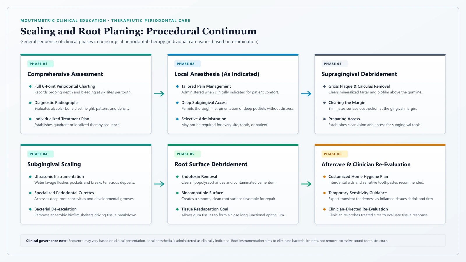
If you have sat in a dental chair and heard numbers like “four,” “five,” or “six millimeters” called out during an examination, you may have been told you need a “deep cleaning.”
This recommendation often brings immediate questions. You may wonder why a routine cleaning is no longer considered sufficient, whether an isolated four-millimeter measurement truly warrants multi-visit therapy, how much discomfort to expect, and whether your dental practice is recommending essential medical treatment or an elective service.
In clinical dentistry, there is no formal procedure called a “deep cleaning.” What dental teams, insurance carriers, and patients colloquially refer to as a deep cleaning is formally designated as periodontal scaling and root planing (SRP). Unlike a routine dental cleaning—a preventive service designed to remove superficial plaque and stains from healthy or mildly inflamed gums—scaling and root planing is a therapeutic medical intervention intended to arrest active periodontitis below the gumline.
Deciding whether scaling and root planing is indicated does not hinge on a single number called out from a chart. A sound clinical recommendation rests on a comprehensive evaluation of your entire periodontium: probing depths, clinical attachment loss, bleeding on probing, subgingival calculus, and radiographic evidence of bone loss. This guide explains what scaling and root planing entails, how clinicians determine when it is considered, what happens during treatment, biological healing, recovery expectations, and dental benefit structures.
MouthMetric Medical Evidence Review
Statements in this guide are grounded in peer-reviewed periodontal literature, consensus clinical practice guidelines from the American Dental Association (ADA) and European Federation of Periodontology (EFP), and landmark histological trials. We do not provide clinical diagnoses or personalized treatment plans. Periodontal health can only be accurately assessed by a licensed dental professional using a calibrated probe and diagnostic radiographs. Nothing here should be construed as reason to decline or delay necessary professional dental therapy. Review the MouthMetric evidence methodology.
What Is a Dental Deep Cleaning?
To understand why a dental team recommends scaling and root planing, it helps to examine the anatomy where tooth meets gum.
In health, gum tissue fits snugly around each tooth. Between the gum margin and tooth surface sits a shallow crevice known as the gingival sulcus, typically measuring 1 to 3 millimeters deep when measured with a calibrated probe. Within this shallow space, daily brushing and flossing effectively disrupt bacterial plaque.
When plaque accumulates along the gumline, chronic inflammation breaks down the connective tissue attachment fibers (the periodontal ligament) anchoring the root into the surrounding jawbone. As these fibers detach, the sulcus deepens into a pathological periodontal pocket.
Subgingival pockets create an oxygen-deprived environment where pathogenic anaerobic bacteria thrive. These species break down proteins into volatile sulfur compounds that frequently produce persistent oral malodor, as detailed in our guide on how to get rid of bad breath. Minerals from saliva and gingival fluid calcify this subgingival plaque into hardened crusts called subgingival calculus, or tartar.
Scaling and Root Planing Explained
Scaling and root planing comprises two distinct yet complementary clinical steps performed beneath the gumline:
- Subgingival Scaling: The mechanical removal of mineralized calculus, bacterial biofilms, and cellular debris from crown and root surfaces extending beneath the gumline into the pocket.
- Root Planing: The deliberate smoothing of rough root surfaces (cementum and superficial dentin) to eliminate embedded bacterial endotoxins (lipopolysaccharides) and necrotic cementum, creating a biologically compatible surface that allows gum tissues to heal and readapt against the root.
Why Subgingival Calculus Requires Professional Instrumentation
Once calculus forms below the gum margin, it cannot be removed with a toothbrush, floss, or home devices. Subgingival calculus bonds tenaciously to root irregularities and acts as a porous reservoir harboring bacterial colonies and cytotoxic byproducts. Leaving these deposits in place perpetuates inflammation and drives progressive bone destruction. Because home care tools cannot safely reach deeper than 2 to 3 millimeters, removing subgingival calculus requires specialized periodontal instruments that access deep pockets without damaging adjacent tissues.
Routine Cleaning vs. Periodontal Treatment: Understanding the Differences
A common source of confusion occurs when someone expects a routine preventive cleaning but is told therapeutic periodontal treatment is necessary.
In the United States, professional dental procedures are classified under the American Dental Association’s (ADA) Current Dental Terminology (CDT) code set. Understanding these categories clarifies why different clinical presentations require distinct treatments.
It is critical to recognize that a CDT code is a procedural and billing classification; it is not a clinical diagnosis, nor does it replace comprehensive clinical judgment.
| Procedure Name | Typical Clinical Presentation | Primary Objective | Target Anatomical Area | CDT Code Identifier |
|---|---|---|---|---|
| Adult Prophylaxis | Clinically healthy gums or localized mild gingivitis; no bone loss | Preventive maintenance; control local irritants | Supragingival tooth surfaces; coronal to the gumline | CDT D1110 |
| Scaling in Generalized Inflammation | Swollen, red, bleeding gums; moderate-to-severe inflammation; no bone loss | Therapeutic reduction of full-mouth gingival swelling | Supragingival and shallow subgingival surfaces | CDT D4346 |
| Full Mouth Debridement | Heavy, dense calculus obscuring teeth and gums; diagnostic evaluation impossible | Preliminary clearing to allow subsequent comprehensive exam | Full mouth; supragingival and gross subgingival deposits | CDT D4355 |
| Periodontal Scaling and Root Planing | Active periodontitis; pocketing, attachment loss, and radiographic bone loss | Therapeutic debridement of infected roots to arrest periodontitis | Subgingival root surfaces within periodontal pockets | CDT D4341 (4+ teeth) & D4342 (1–3 teeth) |
Routine Prophylaxis (CDT D1110)
Routine prophylaxis (CDT D1110) is a preventive procedure for individuals with periodontal health or localized, mild gingivitis. Its clinical objective is removing plaque, calculus, and stains from supragingival tooth surfaces above the gumline. Prophylaxis preserves periodontal health where no bone loss or attachment destruction exists. It is not an appropriate treatment for active periodontitis.
Scaling in Generalized Inflammation (CDT D4346)
Introduced in 2017, CDT code D4346 addresses patients with generalized, moderate-to-severe gingival inflammation (swollen, bleeding gums affecting 30 percent or more of teeth) who have no clinical attachment loss or radiographic bone loss. D4346 provides therapeutic full-mouth scaling to suppress acute gingival inflammation without root instrumentation.
Full Mouth Debridement (CDT D4355)
CDT code D4355 describes a preliminary gross debridement performed when dense calculus encases teeth so extensively that the clinician cannot visualize tooth structures or insert a probe for an accurate exam. Full mouth debridement is not a definitive treatment for periodontitis; it is an initial clearing to enable an accurate diagnosis at a subsequent visit, typically scheduled 2 to 4 weeks later.
Periodontal Scaling and Root Planing (CDT D4341 & D4342)
Periodontal scaling and root planing is therapeutic, indicated specifically when active periodontitis is present—characterized by clinical attachment loss, pocketing, subgingival calculus, and radiographic bone loss.
The CDT code set provides two categories based on disease distribution:
- CDT D4341: Scaling and root planing involving four or more diseased teeth per quadrant, representing comprehensive quadrant therapy.
- CDT D4342: Scaling and root planing involving one to three diseased teeth per quadrant, providing localized therapy so patients with isolated pockets are not billed for or subjected to unnecessary treatment on healthy adjacent teeth.
Why Pocket Depth Alone Does Not Determine Treatment
A pervasive misconception surrounding periodontal care is that a single pocket measurement operates as an automatic trigger for deep cleaning. Patients frequently ask: “My hygienist found a four-millimeter pocket on one molar. Does that automatically mean I need a deep cleaning?”
The evidence-based answer is: no. A pocket depth measurement by itself does not determine whether scaling and root planing is indicated.
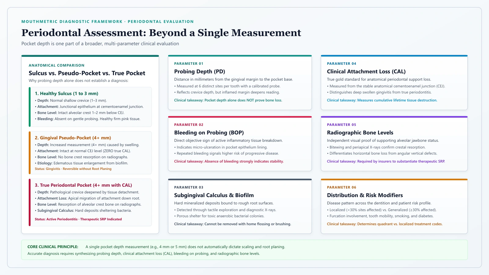
Clinicians evaluate periodontal health through a comprehensive diagnostic synthesis, weighing multiple clinical indicators concurrently:
- Probing Depth (PD): Distance from the gingival margin to the base of the sulcus across six sites per tooth. While 1 to 3 millimeters indicates health, isolated 4-millimeter readings can occur in normal anatomy or mild swelling.
- Clinical Attachment Loss (CAL): Measures whether connective tissue fibers have detached from the fixed anatomical cementoenamel junction (CEJ). As detailed in our guide on gum disease causes and stages, clinical attachment loss is the true hallmark of destructive periodontitis.
- Bleeding on Probing (BOP): Indicates active micro-ulceration of the pocket lining and vascular inflammation. A deep pocket without bleeding may represent a stable, inactive site.
- Radiographic Alveolar Bone Loss (RBL): Diagnostic bitewing and periapical radiographs provide objective evidence of crestal bone resorption.
- Subgingival Calculus and Biofilm: Tactile exploration with an explorer detects mineralized ledges beneath the gumline.
True Periodontal Pockets vs. Inflammatory Pseudo-Pockets
A vital distinction is the difference between a true periodontal pocket and a pseudo-pocket:
- True Periodontal Pocket: Formed by pathological detachment of connective tissue fibers from the root surface and apical migration of the junctional epithelium, accompanied by alveolar bone resorption. True pockets represent structural periodontal destruction.
- Gingival Pseudo-Pocket: Formed when the gingival margin enlarges coronally due to acute edema, hormonal fluctuations, or medication-induced gingival overgrowth, while the underlying connective tissue attachment and bone remain completely intact.
Treating a pseudo-pocket with aggressive root planing is clinically inappropriate because root infection and bone loss are absent. Pseudo-pockets caused by swelling generally resolve with oral hygiene reinforcement and supragingival plaque debridement (such as CDT D4346), not subgingival root instrumentation.
Clinical Decision Framework: When Is SRP Considered?
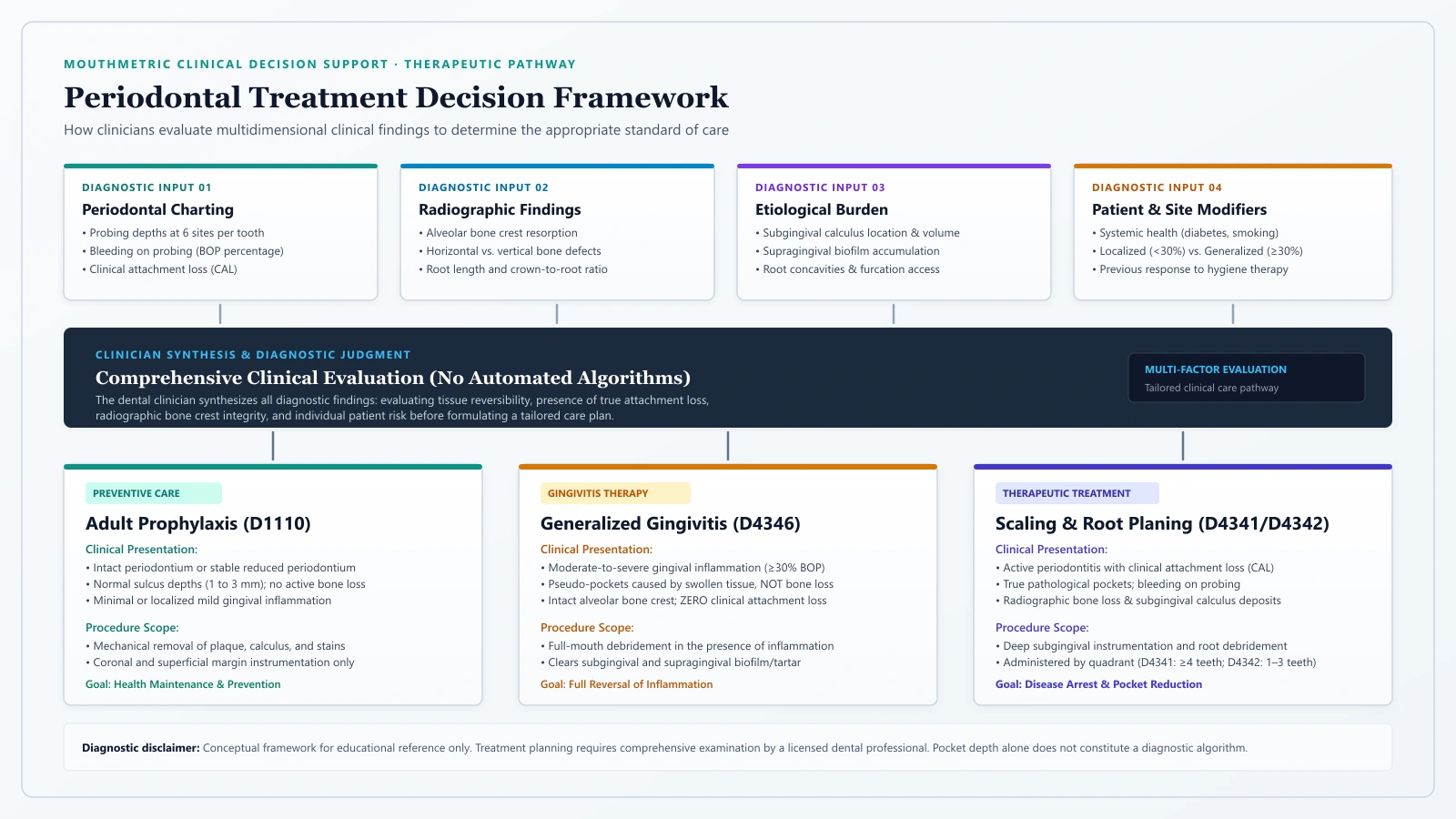
The following framework summarizes how clinical findings guide treatment decisions:
| Clinical Presentation | Probing Depths | Radiographic Bone Loss | Attachment Loss (CAL) | Bleeding on Probing | Evidence-Based Treatment Consideration |
|---|---|---|---|---|---|
| Healthy Periodontium | 1–3 mm | None | None | Absent (< 10% sites) | Routine Prophylaxis (CDT D1110) |
| Gingivitis (Intact Periodontium) | 1–3 mm (or localized 4 mm pseudo-pockets) | None | None | Present (≥ 10% sites) | Routine Prophylaxis (CDT D1110) + Home Care Reinforcement |
| Generalized Severe Gingival Inflammation | 3–4 mm (swollen margins / pseudo-pockets) | None | None | Generalized profuse bleeding | Scaling in Generalized Inflammation (CDT D4346) |
| Localized Mild/Moderate Periodontitis | 4–5 mm at 1 to 3 teeth in a quadrant | Mild crestal bone loss | 1–2 mm localized attachment loss | Present at diseased sites; subgingival calculus detected | Localized Scaling and Root Planing (CDT D4342) |
| Generalized Moderate to Severe Periodontitis | ≥ 4–5 mm across 4+ teeth in a quadrant | Moderate to advanced horizontal or vertical bone loss | ≥ 3–5 mm clinical attachment loss | Persistent bleeding, subgingival calculus, deep pocketing | Quadrant Scaling and Root Planing (CDT D4341) |
As this framework demonstrates, scaling and root planing is reserved for scenarios where clinical attachment loss, radiographic bone loss, and subgingival root contamination exist concurrently. An isolated pocket depth reading, in the absence of attachment loss and bone resorption, does not justify quadrant root planing.
What Happens During a Scaling and Root Planing Procedure
Understanding the practical steps of scaling and root planing helps alleviate dental anxiety and sets clear expectations for care.
Quadrant Scheduling and Appointment Structure
Because scaling and root planing requires meticulous instrumentation beneath the gumline, treating an entire mouth thoroughly in a single visit is rarely practical.
Clinicians typically divide the mouth into four quadrants: Upper Right, Upper Left, Lower Left, and Lower Right. Treatment is commonly scheduled across two separate appointments, addressing one half of the mouth (two quadrants) per visit, with each session lasting 45 to 90 minutes.
Dividing therapy into half-mouth visits limits local anesthesia to one side so you can comfortably eat and speak on the un-numbed side, prevents jaw fatigue, and allows thorough instrumentation of complex pocket anatomies.
Local Anesthesia and Patient Comfort Options
Patient comfort is a primary clinical priority during subgingival instrumentation. Clinicians utilize several anesthetic modalities:
- Topical Numbing Gels: Applied to the mucosa prior to injection to desensitize the surface.
- Needle-Free Subgingival Gels: Thermosetting anesthetic formulations (such as lidocaine/prilocaine combinations) placed directly into pockets with a blunt applicator, providing site-specific soft-tissue anesthesia without lingering lip numbness.
- Local Infiltration and Nerve Blocks: Traditional local anesthetic injections providing complete, profound numbness across teeth, roots, bone, and gingiva.
- Nitrous Oxide or Sedation: Available for patients experiencing heightened dental anxiety.
Ultrasonic Technology and Hand Instrumentation
Modern scaling and root planing combines power-driven ultrasonic scalers with precision manual curettes:
- Ultrasonic Scalers: Utilize high-frequency acoustic vibrations to fracture tenacious calculus off root surfaces rapidly. A stream of cooled irrigation flushes debris out of the pocket while acoustic energy disrupts bacterial cell walls.
- Periodontal Curettes: Specialized hand curettes (such as site-specific Gracey curettes) provide tactile feedback, allowing the clinician to detect microscopic roughness and root concavities, smoothing the root until biologically clean.
Does a Deep Cleaning Hurt? Anesthesia, Sensitivity, and Recovery
A primary concern among patients facing scaling and root planing is procedural comfort and postoperative recovery.
With modern local anesthesia, the procedure itself should not hurt. You will feel gentle pressure, vibrations from the ultrasonic scaler, and water irrigation, but you should not feel sharp pain. If you experience discomfort at any point, alert your clinician immediately so additional anesthetic can be administered.
What Recovery Feels Like: The First 24 to 72 Hours
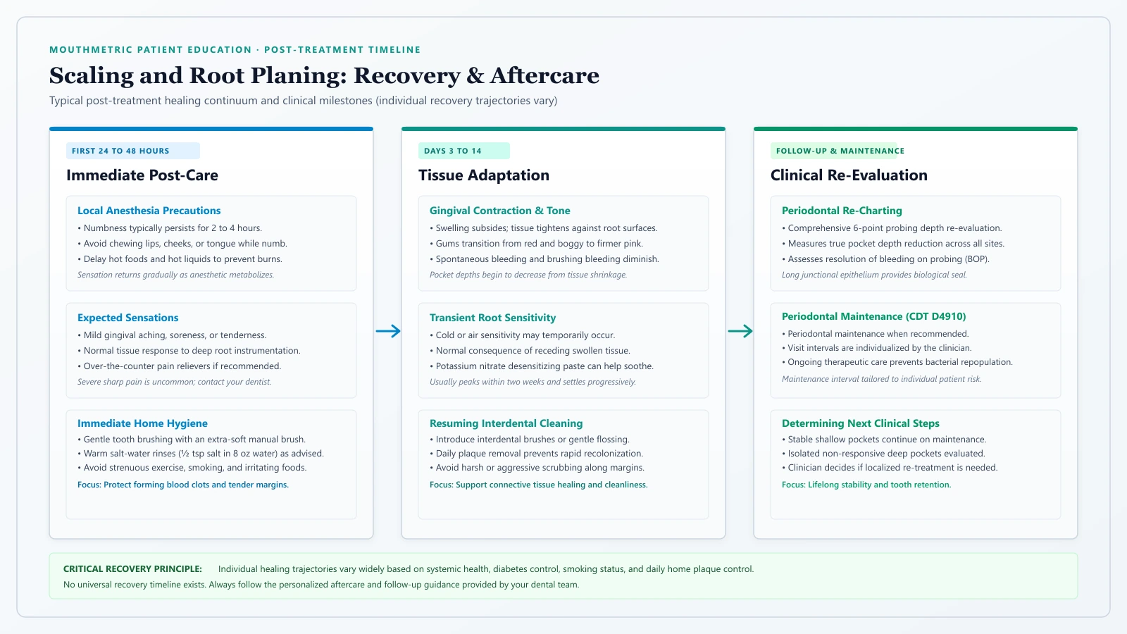
Once local anesthesia wears off—typically within 2 to 4 hours—mild, manageable symptoms are normal:
- Gingival Tenderness: Gums may feel sore or bruised for 24 to 48 hours as mechanical irritation subsides.
- Minor Bleeding: You may notice slight pink-tinged saliva when brushing or rinsing on the evening of the procedure.
- Tooth Mobility Sensation: As thick calculus bridges that previously splinted mobile teeth are removed and inflamed tissues contract, teeth may temporarily feel slightly loose or different to the tongue.
Post-Treatment Comfort Strategies
To support rapid, uneventful healing, dental teams recommend simple home-care strategies:
- Over-the-Counter Analgesics: Mild discomfort is typically managed with anti-inflammatory medications such as ibuprofen (200–400 mg) or acetaminophen, taken according to package instructions.
- Warm Saltwater Rinses: Half a teaspoon of salt dissolved in eight ounces of warm water rinsed gently two to three times daily helps soothe inflamed tissues and promotes healing.
- Gentle Oral Hygiene: Continue brushing twice daily using an ultra-soft toothbrush along the treated gumline. Removing fresh plaque is essential for proper healing.
- Dietary Adjustments: For the first 48 hours, avoid hard, crunchy, sharp foods (chips, nuts, popcorn) that can lodge in healing pockets, as well as highly acidic or heavily spiced foods.
Why Teeth May Feel Sensitive to Cold
Postoperative dentin hypersensitivity to cold liquids and air is a common reaction following scaling and root planing.
This sensitivity occurs due to fluid dynamics within the tooth root, as described by the hydrodynamic theory of dentin sensitivity. When calculus covering the root is removed, microscopic fluid-filled channels called dentinal tubules are exposed to the oral environment. Cold stimuli cause rapid fluid shifts within these tubules, stimulating pulpal nerve fibers.
This sensitivity is almost always temporary. Over several weeks, salivary minerals and minerals from toothpaste occlude exposed tubules, forming a protective surface layer. Using a potassium nitrate or stannous fluoride desensitizing toothpaste accelerates tubule occlusion. If roots remain exposed long term, our guide on gum recession causes and treatments reviews protective varnishes and grafting procedures.
Warning Signs: When to Contact Your Dental Office
While mild soreness is expected, contact your dental practice if you experience severe throbbing pain unresponsive to over-the-counter analgesics, continuous bright red bleeding that persists despite gentle pressure, new facial swelling, discharge of pus, or an elevated body temperature.
How Tissues Heal After Scaling and Root Planing
To understand what scaling and root planing accomplishes, it is necessary to examine the biological processes that occur in healing periodontal tissues.
Tissue Shrinkage vs. True Periodontal Regeneration
An essential concept for patients to grasp is the biological distinction between periodontal repair and periodontal regeneration:
- Periodontal Repair / Healing: The resolution of inflammatory edema, shrinkage of swollen gum margins, and formation of a tight epithelial seal against the root surface. This is what non-surgical scaling and root planing achieves.
- Periodontal Regeneration: The biological recreation of all lost tissues—regrowing new alveolar jawbone, new cementum on the root, and newly oriented periodontal ligament fibers inserting into both bone and root. Non-surgical scaling and root planing does not reliably regrow lost bone.
When scaling and root planing removes subgingival calculus and biofilm, the immune reaction in the pocket wall resolves. Edema subsides, capillaries cease leaking inflammatory fluid, and disorganized inflammatory cells are replaced by healthy collagen fibers.
As a direct consequence of this healing, the swollen gum margin tightens and recedes slightly toward the bone crest. This tissue shrinkage indicates resolving inflammation, though it often makes teeth appear slightly longer and creates small gaps near the gumline.
The Long Junctional Epithelium: How Gums Reattach
Patients frequently wonder whether gums attach back to the tooth root after treatment. In a landmark histological trial, Caton and Zander (1979) demonstrated that healing following scaling and root planing occurs primarily through the formation of a long junctional epithelium (LJE).
Epithelial cells from the gingival margin proliferate downward along the planed root surface, attaching to clean cementum via specialized microscopic adhesion structures called hemidesmosomes.
This long junctional epithelium creates a tight biological seal that prevents bacterial penetration. However, this attachment is an epithelial adhesion, not a new connective tissue attachment anchored into regenerated bone. While a long junctional epithelium maintains clinical health, it remains structurally more fragile than the original developmental connective tissue apparatus, emphasizing the critical need for meticulous long-term plaque control.
Expected Pocket Depth Reductions
Extensive systematic reviews provide clear clinical benchmarks for healing:
- In moderately deep pockets (initially 4 to 6 mm), Cobb (2002) documented an average probing depth reduction of approximately 1.29 mm and a clinical attachment gain of approximately 0.55 mm.
- In deep pockets (initially ≥ 7 mm), average probing depth reduction reaches approximately 2.16 mm, with a clinical attachment gain of approximately 1.19 mm.
These findings were corroborated by Suvan et al. (2020) in a systematic review supporting the European Federation of Periodontology (EFP) S3-level clinical practice guidelines. Their meta-analysis confirmed that subgingival instrumentation produces statistically significant pocket depth reductions across all disease severities.
Approximately half of the observed pocket reduction stems from contraction and shrinkage of the outer gingival margin as swelling resolves; the remaining reduction results from clinical attachment gain via formation of the long junctional epithelium at the base of the pocket.
Why Gums May Look Different: The Reality of “Black Triangles”
Following scaling and root planing, patients may notice small triangular gaps appearing between teeth near the gumline. In clinical dentistry, these spaces are termed open gingival embrasures, colloquially known as “black triangles.”
Patients sometimes assume that the clinician scraped away gum tissue. In reality, the etiology of open embrasures is multifactorial, stemming from pre-existing disease rather than procedural trauma:
- Resolution of Inflammatory Edema: Before treatment, chronically inflamed gums are swollen, fluid-engorged, and thickened. As scaling and root planing eliminates bacterial toxins, swelling subsides and tissue contracts against the underlying bone, unmasking bone loss that had already occurred.
- Interproximal Bone Height: In a landmark study, Tarnow, Magner, and Fletcher (1992) demonstrated that when the distance from the tooth contact point to the bone crest is 5 millimeters or less, the interdental papilla fills the space nearly 100 percent of the time. At 6 millimeters, papilla presence drops to 56 percent; at 7 millimeters or greater, papilla fill occurs in 27 percent or fewer cases.
- Tooth Anatomy: Triangular crowns and divergent roots naturally create wider interdental embrasures when crestal bone resorbs.
Scaling and root planing does not cause black triangles; it reveals the anatomical consequence of historical periodontal bone loss once inflammatory swelling resolves.
The Role of Adjunctive Therapies in Periodontal Care
During a consultation for scaling and root planing, you may be presented with options for adjunctive treatments proposed alongside mechanical debridement. Understanding the evidence behind these options helps you make informed choices.
Locally Delivered Antimicrobials
Locally delivered antimicrobials (LDAs) are antibiotic or antiseptic agents placed directly into individual periodontal pockets, such as minocycline microspheres, doxycycline hyclate gel, or chlorhexidine gluconate chips.
Clinical trials show that local antimicrobials can provide a modest additional pocket depth reduction (typically averaging 0.3 to 0.5 mm) when placed into localized, deep, non-responsive pockets (≥ 5 mm with persistent bleeding). However, clinical practice guidelines from the ADA and EFP emphasize that routine, indiscriminate placement of local antimicrobials across an entire mouth during initial therapy provides limited additional clinical benefit over high-quality mechanical debridement alone.
Systemic Antibiotics: Why Routine Use Is Discouraged
Systemic antibiotics (oral pills such as combinations of amoxicillin and metronidazole) have historically been prescribed alongside scaling and root planing. Today, authoritative clinical guidance has shifted significantly.
The European Federation of Periodontology (EFP) S3-level guidelines (Sanz et al., 2020) and the American Dental Association explicitly discourage the routine use of systemic antibiotics for the vast majority of patients undergoing scaling and root planing. This recommendation is driven by significant global concerns regarding antimicrobial resistance, systemic adverse drug reactions, and gastrointestinal complications.
Systemic antibiotics are not routine adjuncts for most periodontal therapy and may be considered selectively in particular clinical situations—such as severe, rapidly progressing periodontitis in younger individuals or medically compromised patients—based on clinician judgment and patient-specific risk profiles.
Dental Lasers: What Current Evidence Shows
The use of dental lasers (such as diode, Nd:YAG, or Er:YAG lasers) and antimicrobial photodynamic therapy as adjuncts to scaling and root planing has gained widespread commercial popularity.
In a systematic review and meta-analysis of diode lasers as adjuncts to scaling and root planing, Sgolastra et al. (2013) evaluated clinical outcomes across randomized controlled trials. They concluded that while lasers demonstrate bactericidal properties, current scientific evidence demonstrates limited, mixed, or statistically non-significant additional clinically meaningful benefits in pocket depth reduction or attachment gain compared to conventional mechanical debridement alone.
Both ADA and EFP consensus guidelines maintain that mechanical debridement with ultrasonic and hand instruments remains the definitive, evidence-based foundation of periodontal therapy.
Dental Deep Cleaning Costs, Coding, and Insurance Coverage
The financial investment required for scaling and root planing is an understandable concern for patients, especially when unexpected.
Factors Influencing Treatment Fees
Because dental fees in the United States are not federally standardized, out-of-pocket costs vary widely based on several practical variables:
- Geographic Location: Practices in metropolitan areas with high operating overhead typically have higher fee schedules than rural practices.
- Provider Specialty: Care by a board-certified periodontist generally carries higher professional fees than care by a general dentist.
- Extent of Disease (CDT Coding): Treatment fees correspond to the number of diseased teeth per quadrant. Four or more teeth (CDT D4341) carries a higher fee than one to three teeth (CDT D4342).
- Diagnostic Requirements: Initial evaluations often include necessary diagnostic services, such as full-mouth radiographs (FMX) and periodontal charting.
How Dental Benefit Plans Commonly Evaluate Periodontal Claims
Dental insurance coverage for scaling and root planing varies depending on your specific group plan and policy tier.
Unlike routine cleanings—which many dental plans cover at 80 to 100 percent with no deductible—scaling and root planing is almost universally classified as a basic or major periodontal service:
- Deductibles and Co-Insurance: Patients are typically responsible for their annual deductible plus co-insurance, with plans commonly reimbursing 50 to 80 percent of allowable fees.
- Annual Maximum Limitations: Most plans enforce annual benefit caps (commonly $1,000 to $2,000 per benefit year). Full-mouth scaling and root planing can consume a substantial portion of an annual cap.
- Clinical Documentation Requirements: Insurance examiners routinely request supporting clinical documentation before approving benefits. An insurer may request full-mouth radiographs showing crestal bone loss, detailed six-point probing charts, and narrative notes confirming subgingival calculus.
- Frequency Limitations: Many benefit plans restrict scaling and root planing benefits to once every 24 to 36 months per quadrant, with subsequent care reimbursed under periodontal maintenance codes.
Post-Treatment Re-Evaluation and Periodontal Maintenance
Completing scaling and root planing is not the conclusion of periodontal therapy; it marks the transition into long-term disease management.
The Role of Periodontal Re-Evaluation
Following scaling and root planing, soft tissues require time to resolve inflammation and establish a long junctional epithelium. Longitudinal clinical studies show that periodontal tissue healing stabilizes over several weeks.
Dental clinicians typically schedule a formal periodontal re-evaluation several weeks after therapy (most commonly 4 to 8 weeks, with 4 to 6 weeks frequently observed in clinical practice). During this visit, the clinician repeats a complete six-point periodontal probing examination across all treated quadrants to assess pocket depth reductions, resolution of bleeding on probing, tooth mobility changes, and the effectiveness of daily home plaque control.
Sites that have successfully healed (probing depths reducing to ≤ 4 mm with no bleeding) transition into ongoing maintenance. If deep pockets (≥ 6 mm) with persistent bleeding remain, the clinician may discuss localized re-instrumentation, targeted adjunctive care, or referral to a periodontist for surgical periodontal evaluation.
Periodontal Maintenance (CDT D4910): Individualized Long-Term Care
Once a patient has been treated for periodontitis, returning to standard preventive cleanings (CDT D1110) is clinically inappropriate. Periodontitis is a chronic disease; even when pockets heal, the patient remains at elevated risk for disease recurrence.
Ongoing care is provided through Periodontal Maintenance (CDT D4910). Unlike a standard prophylaxis, periodontal maintenance includes continuous monitoring of pocket depths, site-specific subgingival debridement of recurrent pockets, and therapeutic plaque removal.
Recall intervals must be individualized based on disease severity, host risk factors (such as smoking or diabetes), and home care efficacy (Cohen, 2003). A three- to four-month maintenance interval is widely recommended for many post-SRP patients. The biological rationale is supported by research by Sbordone et al. (1990), who demonstrated that in patients with deep periodontal pockets, pathogenic subgingival bacteria repopulate treated root surfaces and return to baseline levels within 9 to 12 weeks following instrumentation if professional debridement is not repeated.
Practical Questions to Ask Your Dental Team
If your dental team has recommended scaling and root planing, open communication ensures that you fully understand your diagnosis. Consider asking these questions:
- “What were my specific probing measurements and where is bone loss visible on my radiographs?” Reviewing your chart helps you visualize areas of active bone resorption.
- “Are my deeper measurements true periodontal pockets with bone loss, or swollen pseudo-pockets?” Confirms whether attachment loss exists or if swelling drives the readings.
- “Do I have bone loss throughout my mouth, or is it confined to specific teeth?” Clarifies whether full-quadrant therapy (D4341) or localized therapy (D4342) is indicated.
- “What comfort options and local anesthetics do you plan to use?” Alleviates procedure-related anxiety.
- “When will we perform my re-evaluation, and what is my recommended maintenance schedule?” Establishes follow-up expectations for long-term disease control.
Common Questions About Deep Cleaning (FAQs)
Is a deep cleaning the same as a regular cleaning?
No. A regular cleaning (CDT D1110) is a preventive service performed above the gumline on healthy or mildly inflamed tissues. Scaling and root planing (CDT D4341/D4342) is a therapeutic procedure performed beneath the gumline to arrest active periodontitis with bone loss and attachment loss.
Does a deep cleaning hurt?
With modern local anesthesia, the procedure itself should not be painful. You will feel vibrations and pressure, but sharp discomfort is prevented by numbing gels, needle-free subgingival anesthetics, or nerve blocks. Mild post-treatment soreness and cold sensitivity are easily managed with over-the-counter pain relievers.
Can a deep cleaning be completed in a single appointment?
While full-mouth single-visit therapy is technically possible, scaling and root planing is most commonly divided into two appointments, treating half the mouth per visit. This prevents bilateral numbness, avoids jaw fatigue, and allows thorough instrumentation.
Will my gums or jawbone grow back after scaling and root planing?
Non-surgical scaling and root planing does not reliably regrow lost alveolar bone. Tissues heal through inflammatory resolution, gum shrinkage, and the formation of a long junctional epithelium along the root surface. True bone regeneration requires surgical regenerative therapies.
Why do spaces (“black triangles”) sometimes appear between teeth after treatment?
Open embrasures (“black triangles”) appear when chronic inflammatory swelling subsides. Resolving edema causes the gum tissue to contract against the remaining bone crest, unmasking pre-existing bone loss.
What happens if I decline scaling and root planing and only get a regular cleaning?
A regular cleaning cannot reach the base of deep periodontal pockets or remove subgingival calculus. Leaving bacterial biofilms undisturbed allows periodontitis to progress silently, leading to progressive bone loss, tooth mobility, and eventual tooth loss.
The Bottom Line
Periodontal scaling and root planing is the clinical gold standard for non-surgical periodontitis management. When recommended based on a comprehensive diagnostic evaluation—integrating probing depths, clinical attachment loss, bleeding on probing, and radiographic bone loss—it effectively arrests destructive gum disease and halts progressive bone resorption.
Healing occurs primarily through tissue shrinkage and the formation of a protective long junctional epithelium. While scaling and root planing does not regrow lost bone, it provides a clean, biologically compatible root surface that allows surrounding tissues to heal. Long-term success ultimately depends on consistent daily plaque control and adherence to individualized periodontal maintenance visits.
Sources and Clinical Evidence
- American Dental Association. CDT 2025/2026: Dental Procedure Codes. Chicago: American Dental Association; 2025.
- Smiley CJ, Tracy SL, Abt E, et al. Evidence-based clinical practice guideline on the nonsurgical treatment of chronic periodontitis by means of scaling and root planing with or without adjuncts. J Am Dent Assoc. 2015;146(7):525-535. PMID: 26113100
- Tonetti MS, Greenwell H, Kornman KS. Staging and grading of periodontitis: Framework and proposal of a new classification and case definition. J Periodontol. 2018;89(Suppl 1):S159-S172. PMID: 29926952
- Caton J, Zander HA. The attachment between tooth and gingival tissues after periodic root planing and soft tissue curettage. J Periodontol. 1979;50(9):462-466. PMID: 114618
- Cobb CM. Clinical significance of non-surgical periodontal therapy: an evidence-based perspective of scaling and root planing. J Clin Periodontol. 2002;29(Suppl 2):6-16. PMID: 12010523
- Cobb CM, Sottosanti JS. A re-evaluation of scaling and root planing. J Periodontol. 2021;92(10):1370-1378. PMID: 33660307
- Cohen RE; Research, Science and Therapy Committee, American Academy of Periodontology. Position paper: Periodontal maintenance. J Periodontol. 2003;74(9):1395-1401. PMID: 14584877
- Sanz M, Herrera D, Kebschull M, et al. Treatment of stage I-III periodontitis—The EFP S3 level clinical practice guideline. J Clin Periodontol. 2020;47(Suppl 22):4-60. PMID: 32383274
- Sbordone L, Ramaglia L, Gulletta E, Iacono V. Recolonization of the subgingival microflora after completion of non-surgical periodontal therapy. J Periodontol. 1990;61(9):579-584. PMID: 2213468
- Sgolastra F, Severino M, Gatto R, Monaco A. Effectiveness of diode laser as adjunctive therapy to scaling root planning in the treatment of chronic periodontitis: a meta-analysis. Lasers Med Sci. 2013;28(5):1393-1402. PMID: 22895576
- Suvan J, Leira Y, Moreno F, et al. Subgingival instrumentation for treatment of periodontitis: A systematic review. J Clin Periodontol. 2020;47(Suppl 22):70-88. PMID: 31889320
- Tarnow DP, Magner AW, Fletcher P. The effect of the distance from the contact point to the crest of bone on the presence or absence of the interproximal dental papilla. J Periodontol. 1992;63(12):995-996. PMID: 1474471